Translate this page into:
Cutaneous larva migrans of the genitalia
Correspondence Address:
Raghavendra Rao
Department of Dermatology, Kasturba Medical College, Manipal-576 104
India
| How to cite this article: Rao R, Prabhu S, Sripathi H. Cutaneous larva migrans of the genitalia. Indian J Dermatol Venereol Leprol 2007;73:270-271 |
 |
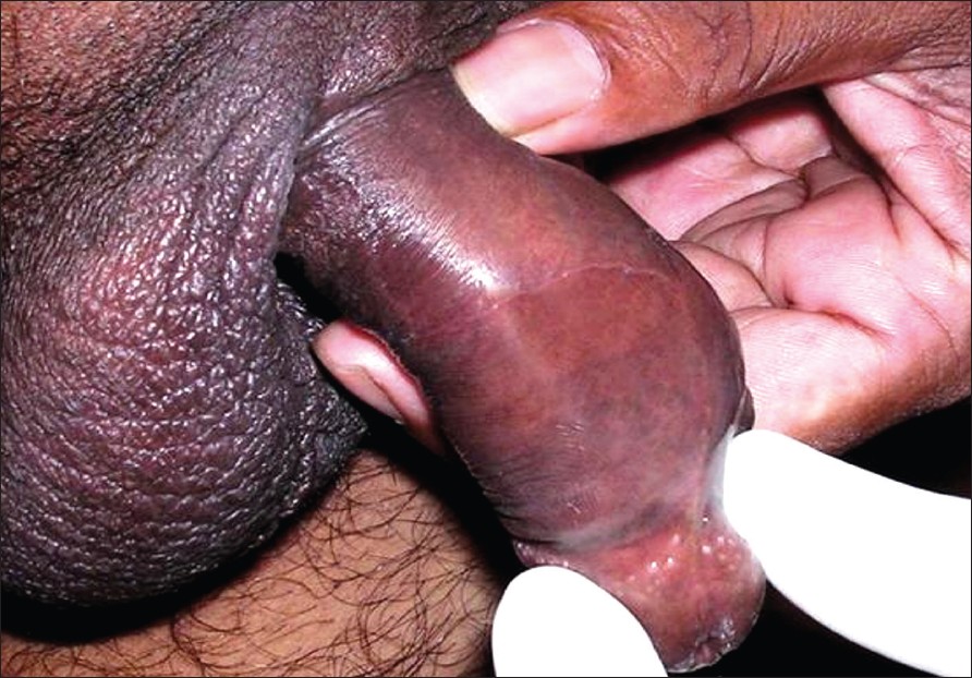
| Figure 1: Serpiginous tract on the shaft of penis |
 |
| Figure 1: Serpiginous tract on the shaft of penis |
Sir,
Cutaneous larva migrans (CLM) is a peculiar dermatitis caused usually by penetration of the skin by hookworm larvae. Common sites of involvement are feet (interdigital spaces, dorsa of feet and the medial aspect of soles), buttocks and hands. We report a case of CLM confined to the penis.
A 35 year-old uncircumcised male presented with an itchy eruption on the penis of four months duration. It started as a small papule on the ventral surface of penis, near the frenulum and subsequently progressed proximally in a serpiginous fashion. He gave a history of a crawling sensation underneath the skin. He denied any history of extramarital sexual exposure. Previous therapies with various topical and systemic antifungals were ineffective.
Cutaneous examination revealed a slightly raised, erythematous, serpentine eruption on the ventral surface of the penis extending from the frenulum to the junction of middle and upper 1/3 rd of the shaft of the penis [Figure - 1]. The distal end was marked by a pearly-white papule. His complete blood count showed eosinophilia and stool examination for parasitic ova and cysts were negative. He was treated with albendazole 400 mg twice daily for three days. Progression of the lesions was halted in three days and complete resolution was seen in a week.
Numerous organisms can cause cutaneous larva migrans (CLM): Ancylostoma brasiliensis , A. caninum , Uncinaria stenocephala and Bubostomum phlebotomum . [1] A. brasiliensis and A. caninum (the dog and cat hookworms) are the most common causes. Most of the larvae are unable to undergo further development in humans (accidental host) and die within 2-8 weeks time. [2] Though the condition is worldwide in distribution, it is substantially more common in tropical and subtropical countries. Activities that increase the risk of infestation include walking barefoot on a beach, working in the garden and playing in sandpits. The incubation period varies between 1-6 days. The clinical features of CLM vary from nonspecific dermatitis at the site of penetration of the larva to a typical creeping eruption.
After penetration, the larva can lie quiescent for weeks or immediately begin their creeping activity. The characteristic lesion of CLM consists of slightly raised, erythematous thread-like linear or serpentine tracks. The condition is extremely itchy. Large number of larvae may be active at the same time with the formation of a disorganized series of loops and tracks. The larva usually lies somewhat in front of the head of the track. Vesiculobullous lesions along the tracks and folliculitis are other uncommon manifestations. [3],[4] Excoriation and impetiginization of the lesion are common.
CLM confined to the penis is very rare with the mode of larval entry being unclear in such cases. Our patient hails from the coastal area and used to spend his leisure time on the beach. Karthikeyan et al have speculated that the habit of not wearing any underwear while playing on the beach is a possible cause of such penetration. [5] This could be applicable to our patient too.
Skin biopsy is of little help and the diagnosis is mainly clinical. Epiluminescence microscopy is a noninvasive method to detect larva and confirm diagnosis. [6] Differential diagnosis of CLM includes cercarial dermatitis, migratory myiasis and contact dermatitis. Surgery and cryotherapy are ineffective as the larva is easily missed, being ahead of the visible track. A single dose of Ivermectin (150-200 µg/kg) is the best treatment. Albendazole (400-800 mg/day) for three days and topical thiabendazole (10%) are also useful.
| 1. |
Vega-Lopez, Hay RJ. Parasitic worms and protozoa. In : Rook's Text book of Dermatology. Burns T, Breathnach S, Cox N, Griffith C, editors. 7 th ed. Oxford: Blackwell Science; 2004. p. 32.1-32.48.
th ed. Oxford: Blackwell Science; 2004. p. 32.1-32.48.'>[Google Scholar]
|
| 2. |
Padmavathy L, Rao LL. Cutaneous larva migrans- A case report. Indian J Med Microbiol 2005; 23:135-6.
[Google Scholar]
|
| 3. |
Veraldi S, Arancio L. Giant bullous cutaneous larva migrans. Clin Exp Dermatol 2006;31:613-4.
[Google Scholar]
|
| 4. |
Veraldi S, Bottini S, Carreca C, Gianotti R. Cutaneous larva migrans with folliculitis: A new clinical presentation of the infestation. J Eur Acad Dermatol Venereol 2005;19:628-30.
[Google Scholar]
|
| 5. |
Karthikeyan K, Thappa DM, Jeevan Kumar B. Cutaneous larva migrans of the penis. Sex Trans Infect 2003;79:500.
[Google Scholar]
|
| 6. |
Eisher E, Themes M, Worret WC. Cutaneous larva migrans detected by epiluminescent microscopy. Acta Derm Venereol 1997;77:487-8.
[Google Scholar]
|
Fulltext Views
5,216
PDF downloads
1,788


