Translate this page into:
Cutaneous neonatal lupus erythematosus
Correspondence Address:
Ane Jaka
Servicio de Dermatolog�a, Hospital Donostia, Paseo Dr. Begiristain, Donostia-San Sebasti�n, (Gipuzkoa)
Spain
| How to cite this article: Jaka A, Zubizarreta J, Ormaechea N, Tuneu A. Cutaneous neonatal lupus erythematosus. Indian J Dermatol Venereol Leprol 2012;78:775 |
Sir,
Neonatal lupus erythematosus (NLE) is caused by transplacental passage of maternal anti-Ro and/or anti-La autoantibodies. The most common manifestations are cutaneous lupus lesions, third-degree heart block, and/or hematologic cytopenias. The disease is a transient process, although complete heart block, once established, is permanent. [1]
We describe an eight-month child with annular erythematous cutaneous lesions on the face and a history of a brother died of heart block, which are the major organs affected in neonatal lupus erythematosus.
A Caucasian eight-month child presented with a one-month history of cutaneous lesions on the face. During the pregnancy the mother was treated with corticosteroid because she previously had two abortions and a newborn died because of complete heart block.
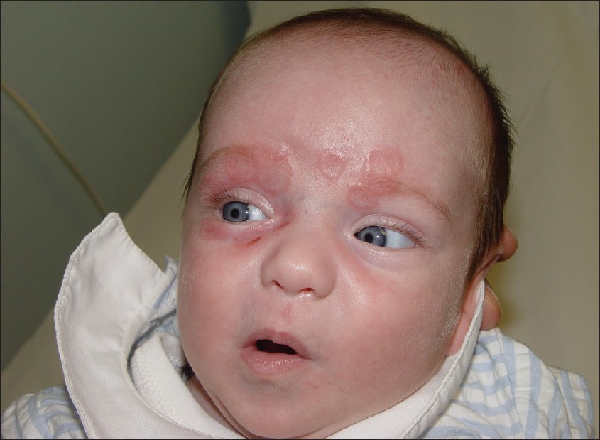
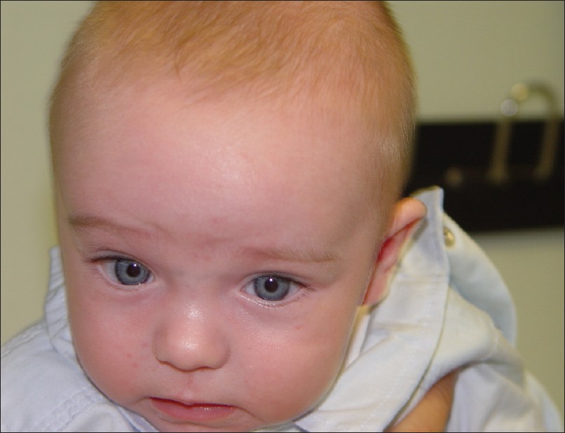
Physical cutaneous examination revealed erythematous annular plaques with central atrophy confluent in periorbital area [Figure - 1]. The remainder of the cutaneous examination was not remarkable. Laboratory studies revealed positive ANA and anti-Ro/anti-La. Blood count, liver enzymes and cardiologic examination were normal, including echocardiography. Maternal serum was also positive for antinuclear antibodies and anti-SSA/Ro at the time we suspected the diagnosis of NLE in the child, but she was not diagnosed with any connective disease. Cutaneous neonatal lupus erythematosus diagnosis was made based on the characteristic skin changes, laboratory findings and maternal history. Photoprotection was prescribed and within a few months the cutaneous lupus lesions resolved spontaneously [Figure - 2] and the serum antibodies disappeared.
 |
| Figure 1: Erythematous annular plaques with central atrophy, confluent in periorbicular area |
 |
| Figure 2: 1 month later, after spontaneous resolution of the cutaneous lupus lesions |
NLE is an uncommon autoimmune disease caused by transplacental passage of maternal anti-Ro and/or anti-La autoantibodies, but it has not been proven unequivocally that maternal autoantibodies cause the disease. It seems to be more determinants of clinical expression of the disease. Several studies have demonstrated the importance of genetic factors and described an increased prevalence of HLA-DR3 in mothers of children affected by NLE. Moreover, it has been observed a major HLA-DR2 is more frequent in healthy children of mothers with anti-Ro antibodies and their mothers, when compared with children affected by NLE and healthy population. [2] However, there are probably environmental factors that also contribute to the expression of this disease.
The most common clinical findings include cutaneous lesions and third-degree heart block. [3] Hepatic abnormalities or hematologic cytopenias are less frequently reported. Most often, only one of these organ systems is affected, but involvement of two or more is also possible.
The cutaneous findings are consistent with cutaneous lupus and most similar to the subacute cutaneous lupus erythematosus subtype, [4] affecting the face or scalp, but they are also common on the trunk and extremities. Less frequently cutaneous lesions presented as a confluent erythema around the eyes, giving an "eye-mask" appearance. [5] Skin lesions are often present at the first few weeks of life, but may be present at birth. They usually have photosensitivity and a low risk of scarring.
The diagnosis of cutaneous NLE is made with the clinical examination and maternal history, with verification of the presence of maternal anti-Ro autoantibodies.
Neonatal lupus disease process is transient and cutaneous lesions resolve completely within weeks or few months. Otherwise, complete heart block, once established, is permanent. [4] This is a common and severe manifestation of neonatal lupus and the majority of cases require pacemaker implantation. There are studies that indicate that treatment of anti-Ro-positive mothers with corticosteroid early in pregnancy may prevent heart block. [5]
Children who have had neonatal lupus can have an increased risk for autoimmune diseases later in the childhood, because they have a family history of autoimmunity. Mothers may have systemic lupus erythematosus, Sjögren syndrome, or another connective tissue disease, or may not have any of them [3] (only autoantibodies in serum).
In children under suspicion of neonatal lupus, laboratory studies with serum autoantibodies and blood count with hepatic enzyme, as well as a cardiologic examination should be performed.
| 1. |
Wisuthsarewong W, Soongswang J, Chantorn R. Neonatal lupus erythematosus: Clinical character, investigation, and outcome. Pediatr Dermatol 2011;28:115-21.
[Google Scholar]
|
| 2. |
García-F-Villalta MJ, Torrelo A, Mediero IG, Zambrano A. Lupus eritematoso neonatal en dos hermanas gemelas. Actas Dermosifiliogr 2003;94:313-5.
[Google Scholar]
|
| 3. |
Lee LA.Cutaneous lupus in infancy and childhood. Lupus 2010;19:1112-7.
[Google Scholar]
|
| 4. |
Requena C, Pardo J, Febrer I. Lupus eritematoso infantil. Actas Dermosifiliogr 2004;95:203-12.
[Google Scholar]
|
| 5. |
Lee LA. Transient autoimmunity related to maternal autoantibodies: Neonatal lupus. Autoimmun Rev 2005;4:207- 13.
[Google Scholar]
|
Fulltext Views
3,651
PDF downloads
3,601


