Translate this page into:
Cutaneous sarcoidosis: Clinical profile of 23 Indian patients
Correspondence Address:
Nand Lal Sharma
Department of Dermatology, Venereology and Leprosy, Indira Gandhi Medical College, Shimla - 171 001 (H.P.)
India
| How to cite this article: Mahajan VK, Sharma NL, Sharma RC, Sharma VC. Cutaneous sarcoidosis: Clinical profile of 23 Indian patients. Indian J Dermatol Venereol Leprol 2007;73:16-21 |
Abstract
Background: Sarcoidosis is a multisystem disease of undetermined etiology. Indian studies on cutaneous sarcoidosis are not many and mainly comprise case reports. Aims: This retrospective study was carried out to assess the clinical profile of sarcoidosis patients presenting with cutaneous lesions. Methods: All histopathologically proven cases of cutaneous sarcoidosis seen consecutively between 1999 and 2004 were studied. Their age, sex, presenting features, evolution of disease and laboratory parameters were analyzed. Results: A total of 23 patients (F:M 15:8) between 31 to 78 years (mean 44.3 years) of age had the mean duration of skin lesions of 1.4 years. Six patients had one to four lesions; two patients each had scar sarcoidosis and angiolupoid and one patient each had recurrent erythema nodosum, leg lymphedema and subcutaneous sarcoidosis. Others showed combination of papules, nodules, plaques and psoriasiform lesions. Peripheral lymph nodes were involved in two patients. Among 10 patients of pulmonary involvement, three had become symptomatic four months to four years after the cutaneous lesions. Routine laboratory investigations including serum calcium estimation were normal in all cases. Serum angiotensin-converting enzyme levels were raised in 3 out of 6 patients. Asymptomatic lytic lesions of digital bones were detected in hand X-ray of one patient.Conclusion: Skin lesions of sarcoidosis are like the tip of an iceberg indicating more changes in other organs. The symptomatology and abnormal laboratory results do not necessarily correlate with the severity of cutaneous involvement in general.


Introduction
Sarcoidosis, a multisystem disease of obscure etiology, is characterized by the formation of noncaseating epithelioid cell granulomas in several organs or tissues. The main organs affected are the lungs, lymph nodes and eyes in the order of frequency. Cutaneous involvement is polymorphic and is seen in 20-35% patients with sarcoidosis.[1],[2] Out of these cases, about 25% patients have exclusively cutaneous affection.[3]
The incidence of sarcoidosis varies among countries and different ethnic groups.[4],[5] The recorded prevalence is 10 and 40 per 100000 population in the US and Europe respectively.[4] In India, the exact prevalence of sarcoidosis is not known. It has been variously estimated as 61 and 150 cases per 100000 outdoor patients in Delhi and Kolkata hospitals respectively in the past.[6] The reporting of cases, particularly of cutaneous sarcoidosis, is also very low and mainly comprises single case reports. We studied the clinical profile of 23 cases of cutaneous sarcoidosis encountered by us during a span of five years.
Methods
Twenty-three patients of cutaneous sarcoidosis detected consecutively during 1999-2004 were studied. All these patients had presented on their own, primarily for their cutaneous lesions. The diagnosis was mainly on clinical suspicion and confirmed by histopathologic examination of at least one skin lesion. They were subjected to detailed history and thorough dermatologic, ophthalmic and systemic examination. Patients having perinostril or perioral lesions were particularly screened for upper airway involvement. Chest X-rays and spirometric pulmonary function tests, routine hematological investigations, hepato-renal assessment, serum glucose, electrolytes and calcium estimations were performed in all of them. In addition other relevant investigations were performed in variable number of patients. Results of radiographic investigations and spirometry were interpreted in consultation with respective consultants.
Results
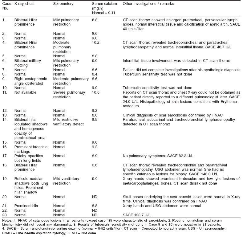
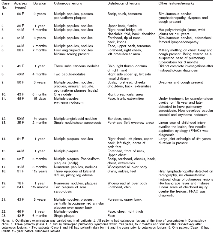
The age, sex, duration of disease, number and morphology of cutaneous lesions, systemic features and investigative profile are tabulated [Table - 1],[Table - 2]. These 23 patients were between 31 to 78 years of age (mean 44.3 years). There were 15 women (31-78 years) and eight men (40-50 years) having a ratio 15:8. There were no children.
Disease profile
A combination of numerous papular, nodular and plaque lesions was a commoner presentation as compared to six patients having one to four lesions. The lesions were widely distributed over the face, trunk and extremities with a mean duration of 1.4 years.
One patient each had recurrent episodes of fever, arthralgia, erythema nodosum (EN) and diffuse pitting edema over shins, ankles and feet of 15 days and 1½ years duration respectively. The presenting features in the other 21 patients were polymorphic skin lesions of one and a half months to nine years duration. The lesions were asymptomatic and variable hue of lesional erythema was characteristic.
Two patients had annular plaques, involving the face, scalp and forearm in one and the back in the other. A few psoriasiform plaques, particularly over the scalp, were present in three patients in addition to multiple lesions of different morphology. Two patients had angiolupoid noduloplaques over the face, scalp and ear lobes. Subcutaneous nodules of sarcoidosis were seen over the right hand in one patient. Two patients presented with sarcoid nodules fixed to overlying scars of childhood injuries over the forehead and chin.
Polyarthralgia involving the ankles, knees and elbows, one and a half to four and a half years before onset of cutaneous lesions was complained of by two patients, while another patient had history of episodes of low-grade fever and malaise associated with numerous sarcoidal plaques. One patient had been treated for granulomatous uveitis one and a half year prior to onset of multiple papular lesions of sarcoidosis, erythema nodosum and other features of L φfgren′s syndrome. Cervical and cervico-femoral lymphadenopathy was found in two separate patients. Three patients developed shortness of breath and cough four months to four years after the cutaneous lesions had appeared. Ten patients showed simultaneous involvement of lungs or pleura documented by X-ray, spirometery and/or CT thorax.
Histopathologic features
Lesional skin biopsies in 20 patients and fine needle aspiration cytology of scar sarcoid lesions in two patients showed characteristic multiple sarcoidal granulomas. Occasional specimen also had fibrinoid necrosis, multinucleated giant cells and asteroid or Schaumann bodies. Histopathology of involved lymph nodes in two of these patients too had granulomatous pathology consistent with sarcoidosis. The only patient having lymphedema of legs had no specific skin lesion that could be subjected to biopsy.
Radiologic features
Chest skiagrams of 10 patients had findings suggestive of pulmonary sarcoidosis. Six of these patients had bilateral prominent hilar shadows while bilateral miliary mottling, reticulo-nodular shadows with hilar prominence, patchy opacities and prominent bronchial markings were seen in one patient each. Obliteration of right costophrenic angle suggesting minimal pleural effusion was noted in one case. Four patients with bilateral hilar prominence showed enlarged pre- and para-tracheal, tracheobrancheal, subcarinal groups of mediastinal lymph nodes in thoracic computed tomographic (CT) scans.
The patient having leg lymphedema, though showed mediastinal lymphadenopathy, had no associated para-aortic lymphadenopathy on abdominal ultrasonography. Skull X-ray showed no bone abnormality underlying scar sarcoid lesions. Prominent trabeculae and a few lytic lesions of metacarpo-phalangeal bones were seen in hand X-ray of a 78-year-old patient having numerous cutaneous lesions and extensive nodulo-reticular pulmonary shadows in chest skiagram. She otherwise had no respiratory symptoms.
Investigative profile
Serum angiotensin-converting enzyme levels were above normal in three of six patients. The only patient having granulomatous uveitis (L φfgren′s syndrome) and generalized papular lesions had associated severe restrictive airway disease while mild to moderate restrictive airway abnormality was recorded in the spirometery of six other patients.
None of the patients had significant abnormalities of routine hematology, serum biochemistry and urinalysis. Results of tuberculin sensitivity available in 21 patients were all negative. Two patients did not complete investigations after histopathologic diagnosis.
Discussion
Sarcoidosis affects both the sexes with a slight preponderance among females. Most patients present between 20-40 years of age but the disease can affect children and the elderly as well.[4] Females outnumbered males by almost twice in this report. The majority (22) of the patients were between 31-52 years. The oldest patient was a 78-year-old female.
In a recent review, 73% patients had only cutaneous lesions at the beginning of their disease. About 70% of them had systemic manifestations concomitantly and the remaining 30% patients developed systemic disease between six months to three years thereafter.[2] Our three patients developed pulmonary symptoms four months to four years after the cutaneous lesions and one patient had ocular involvement (uveitis) one and a half year prior to cutaneous lesions. The onset of the disease is usually insidious but may be occasionally associated with lethargy, weight loss and general malaise.[7] Polyarthritis and moderate to high fever, however, are usual in patients presenting with erythema nodosum. Only one patient, a 78-year-old female, had history of low-grade fever, malaise and numerous widespread cutaneous lesions. Two other patients having episodic fever and polyarthralgia had associated erythema nodosum and leg lymphedema.
A strong clinical suspicion and characteristic histopathology is important for the diagnosis. The polymorphic lesions usually exhibit a mixed pattern and distribution[8] and were also evident in our 22 patients. Angiolupoid lesions, seen in only two patients, are rare. They are soft, hemispherical and have orange-red or reddish-brown color due to marked telangiectatic component.[9] Annular lesions seen in the chronic stage (>2 years) result from peripheral evolution and central clearing with hypopigmentation, atrophy or scarring.[7] They have a predilection for the face, forehead and neck. Two patients with sarcoidosis of about three years duration had characteristic annular and arcuate lesions. Their presence over the back and forearm is uncommon.
In papular sarcoid, lesions are small, 1-5 mm in size and occur in hundreds. Initial orange or yellow-brown color turns brownish red or violaceous and involutes later to faint macules. In general they are associated with more favorable prognosis.[2] However, our sole patient having predominantly papular lesions had severe pulmonary involvement and had taken treatment for granulomatous uveitis in the past indicating a more progressive disease.
Subcutaneous nodular lesions (Darier-Roussy sarcoid) occur in < 5% of sarcoidosis patients. They are characterized by 1-3 cm-sized deep-seated nodules having violaceous overlying epidermis, localized mainly over the trunk or extremities and have no predictive value.[10] Biopsy is needed for diagnosis. Our patient of subcutaneous sarcoidosis did not complete other investigations after histopathologic confirmations of the diagnosis.
Development of sarcoidosis lesions on scars is a specific but uncommon manifestation.[11] Previously silent scars get infiltrated, become erythematous or violaceous but remain asymptomatic and represent a benign disease. Two of our patients had sarcoidosis developing in very old scars without any systemic involvement. Diagnosis of these lesions by fine needle aspiration cytology appears possible with reasonable accuracy.
Lupus pernio, a rare form of cutaneous sarcoidosis, was once thought to occur more commonly in patients of European origin.[12] Lesions may be disfiguring and often associated with chronic fibrotic involvement of respiratory tract and osteolytic lesions of digital bones. They rarely involute spontaneously. It has not been reported from India previously[6] and was also not seen in any of our patients.
Reports on the frequency of EN in sarcoidosis are variable.[12] L φfgren′s syndrome appears to be a more complete form of this reactive variety. In a large series of 251 cases of EN, it indicates good prognosis as> 80% patients showed spontaneous resolution of sarcoidosis in < 2 years.[13] As a general rule, specific lesions and not EN, are associated with more progressive disease.[14] Severe pulmonary disease was observed in our only patient having L φfgren′s syndrome.
Bilateral but asymmetric leg lymphedema as seen in one patient in the present report is a very rare manifestation.[12] It occurs due to involvement of lymph nodes in the iliac or para-aortic region. Our patient though found having mediastinal lymphadenopathy, did not show abdominal lymphadenopathy probably due to early stage of the disease and appeared to be nonprogressive during the year of follow-up.
No organ system appears to be exempt from occasional involvement of sarcoidal granulomas. Granulomatous pulmonary involvement occurs in over 90% cases followed by sarcoidosis of intrathoracic and peripheral lymph nodes in about 75-90% patients [4],[15] Irreversible lung fibrosis and severe disability occurs in 10-15% patients.[7] Ophthalmic involvement manifests in about 25% patients as anterior or posterior uveitis, conjunctival nodule and lacrymal gland and duct lesions, occurring in that order.[16] Pulmonary/pleural involvement, asymptomatic or of variable severity in 10 patients, lymph nodes involvement in two and uveitis in one patient of the present series conform to this well established form of systemic sarcoidosis. Polyarthralgias occur with acute sarcoidosis or as a component of chronic disease[10] and have been observed in two patients in this series. They had it for one and a half to four and a half years preceding the appearance of cutaneous lesions sans systemic involvement. The liver is often affected, manifesting as biochemical abnormality in 20-30% patients.[4] Renal involvement with increased calciuria, with or without hypercalcemia, is uncommon.[4] None of our patients appeared to have involvement of the hepatorenal system.
Chest X-ray findings usually fall in one of the three patterns - bilateral hilar and mediastinal lymphadenopathy, diffuse parenchymal involvement and parenchymal involvement with hilar lymphadenopathy.[4] Variable parenchymal involvement and/or hilar or mediastinal lymphadenopathy was discovered in nine patients in this series on chest skiagrams or thoracic CT scans. Infiltration of pleura and pleural effusion, that may be massive sometimes, have been documented rarely.[6],[17] However, pleural effusion is more often unilateral, small and detected on sonography,[6] as seen in one of our patients who had blunting of a costophrenic angle due to minimal pleural effusion. Lytic lesions of hand bones observed in one of our patients with extensive cutaneous lesions have been well described previously. Asymptomatic bone changes, especially lysis with bone cysts involving small bones of hands and feet are more common and destructive in black South African patients[13] but are uncommon in Indians.[6],[12]
Elevated serum angiotensin-converting enzyme level, seen in 50-60% patients, is neither a sensitive nor a specific diagnostic marker of sarcoidosis and does not reliably correlate with disease activity or its prognosis.[7],[18] Negative tuberculin sensitivity is a well known feature of sarcoidosis and extremely important in tuberculosis endemic areas like ours. All those patients tested for tuberculin sensitivity in this report had negative reaction.
Delayed diagnosis appears to be another feature in this part of the world where tuberculosis remains the first diagnostic possibility. Anti-tuberculosis treatment is often given for prolonged periods before the diagnosis of sarcoidosis is finally made. High index of clinical suspicion therefore is of paramount importance.
The prognosis of patients with cutaneous sarcoidosis depends mainly on the extent of systemic involvement. However, the extent of cutaneous lesions does not correlate with that of systemic involvement and their prognostic value remains unclear.[19] The present report also reflects no correlation between cutaneous involvement and systemic features or abnormal laboratory results in general.
Acknowledgments
Departments of Ophthalmology and Pulmonary Medicine, I. G. Medical College, Shimla, Himachal Pradesh provided their valuable help and guidance in carrying out this study.
| 1. |
Kerdel FA, Moschella SL. Sarcoidosis. An updated review. J Am Acad Dermatol 1984;11:1-19.
[Google Scholar]
|
| 2. |
Mana J, Marcoval J, Graells J, Salazar A, Peyri J, Pujol R. Cutaneous involvement in sarcoidosis: Relationship to systemic disease. Arch Dermatol 1997;133:882-8.
[Google Scholar]
|
| 3. |
Hanno R, Needelman A, Eiferman RA, Callen JP. Cutaneous sarcoidal granulomas and the development of systemic sarcoidosis. Arch Dermatol 1981:117:203-7.
[Google Scholar]
|
| 4. |
Braverman IM. Sarcoidosis. In : Feedberg IM, Eisen AZ, Wolff K, Austen KF, Goldsmith LA, Katz SI, editors. Fitzpatrick's Dermatology in General Medicine. 6th ed. Mcgraw Hill: New York; 2003. p. 1777-83.
[Google Scholar]
|
| 5. |
Edmondstone WM, Wilson AG. Sarcoidosis in Caucasians, Blacks and Asians in London. Br J Dis Chest 1985;79:27-36.
[Google Scholar]
|
| 6. |
Gupta SK. Sarcoidosis: The Indian scene. Indian J Clin Pract 1993;3:19-27.
[Google Scholar]
|
| 7. |
English JC, Patel PJ, Greer KE. Sarcoidosis. J Am Acad Dermatol 2001;44:725-43.
[Google Scholar]
|
| 8. |
Okano M, Nishimura H, Morimoto Y, Maeda H. Faint erythema. Another manifestation of cutaneous sarcoidosis? Int J Dermatol 1997;36:681-4.
[Google Scholar]
|
| 9. |
Gawkrodger DJ. Sarcoidosis. In : Champion RH, Burton JL, Burns DA, Breathnach SM, editors. Textbook of Dermatology. 6th ed. Blackwell Science: Oxford; 1998. p. 2679-700.
[Google Scholar]
|
| 10. |
Odom RB, James WD, Berger TG. Sarcoidosis. In : Odom RB, James WD, Berger TG, editors. Andrews' Diseases of the skin. 9th ed. WB Saunders Co: Philadelphia; 2000. p. 896-910.
[Google Scholar]
|
| 11. |
Nayar M. Sarcoidosis on ritual scarification. Int J Dermatol 1993;32:116-8.
[Google Scholar]
|
| 12. |
Jacyk WK. Cutaneaous sarcoidosis in black South Africans. Int J Dermatol 1999;38:841-5.
[Google Scholar]
|
| 13. |
Neville E, Walker AN, James DG. Prognostic factors predicting the outcome of sarcoidosis: An analysis of 818 patients. Q J Med 1983;52:525-33.
[Google Scholar]
|
| 14. |
Veien NK, Stahl D, Brodthagen H. Cutaneous sarcoidosis in Caucasians. J Am Acad Dermatol 1987;16:534-40.
[Google Scholar]
|
| 15. |
Lynch JP 3rd, Kazerooni EA, Gay SE. Pulmonary sarcoidosis. Clin Chest Med 1997;18:755-85.
[Google Scholar]
|
| 16. |
Ghabrial R, McCluskey FJ, Wakefield D. Spectrum of sarcoidosis involving the eye and brain. Aus NZJ Opthalmol 1997;25:221-4.
[Google Scholar]
|
| 17. |
Warshawsky ME, Shanies HM, Rozo A. Sarcoidosis involving the thyroid and pleura. Sarcoidosis Vasc Diffuse Lung Dis 1997;14:165-8.
[Google Scholar]
|
| 18. |
Callen JP, Hanno R. Serum angiotensin-converting enzyme levels in patients with cutaneous sarcoidal granulomas. Arch Dermatol 1982;2:232-3.
[Google Scholar]
|
| 19. |
Minus HR, Grimes PE. Cutaneous manifestations of sarcoidosis in Blacks. Cutis 1983;32:361-3.
[Google Scholar]
|
Fulltext Views
5,938
PDF downloads
4,299


