
Translate this page into:
Cutaneous signs in infectious diseases
Corresponding author: Dr. Ashish Amrani, Department of Dermatology, NC Medical College and Hospital, Panipat, Haryana, India. ashishamrani@gmail.com
-
Received: ,
Accepted: ,
How to cite this article: Amrani A, Sil A, Das A. Cutaneous signs in infectious diseases. Indian J Dermatol Venereol Leprol 2022;88:569-75.
A “sign” refers to the objective finding, which a clinician observes during the clinical examination of the patient. Signs implicate significant changes or abnormal findings which can assist the clinician in making the final diagnosis of any disorder. Cutaneous signs indicate various dermatological and systemic disorders and sometimes help the dermatologist in making a spot diagnosis. Various cutaneous signs have been described in many infectious disorders. Here, an attempt is made to compile various such cutaneous signs seen in infectious diseases.
Apple jelly sign: It denotes yellowish-brown granulomatous appearance seen on diascopy, resembling apple jelly and indicates collection of tubercles in dermis with degenerative changes. They are characteristically seen in lupus vulgaris, and also been described in sarcoidosis and lupoid cutaneous leishmaniasis [Figure 1].1
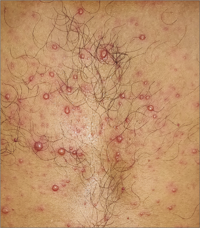
Blueberry muffin sign (Blueberry muffin baby or rash): Neonatal eruption characterized by non-blanchable violet-red papulo-nodules due to extramedullary (dermal) erythropoiesis. They are typically seen in infection with toxoplasma, rubella, cytomegalovirus and herpes simplex virus. Hematological dyscrasias, neonatal neuroblastoma, congenital alveolar rhabdomyosarcoma, neonatal lupus erythematosus and congenital Langerhans cell histiocytosis are the non-infectious conditions where this sign may be seen[Figure 2].2
BOTE sign: In molluscum contagiosum, signs of inflammation (tenderness, erythema and crusting) represent a robust host response that often precedes the resolution of the viral disease. The acronym “BOTE sign” (beginning of the end) has been proposed to help underscore the significance of the inflamed lesions as an expected variation in the evolution of immune response to the virus, rather than bacterial superinfection [Figure 3].3,4
Breakfast, lunch and dinner sign: The bites of bed bugs (Cimex lectularius) usually follow a linear pathway in a group of three to five blood meals and are often referred to as “breakfast, lunch and dinner” or “breakfast, lunch and supper” sign [Figure 4].5
Calabar sign: Also known as Calabar swellings, they are transient subcutaneous swellings, marking the migratory course of the adult filarial eye worm, Loa loa through the tissues.6
Cayenne pepper pus sign: This sign indicates actinomycosis where cayenne pepper granules are observed with drops of pus.7
Chik sign: Centro-facial distribution of pigmentation, especially involving the nose is one of the cutaneous manifestations of chikungunya fever.8,9 Recently this sign has also been observed in dengue fever [Figure 5].10
Comby sign: It is an initial sign of measles, comprising thin whitish patches on the gingiva and buccal mucosa, made up of desquamating epithelial cells.7
Comet sign: Dermatitis caused by Pyemotes species (a parasite of the furniture beetle) presents as erythematous pruritic macules or urticated plaques surmounted by vesicles; occasionally they may be bullous. These lesions are sometimes associated with a specific linear erythematous macular tract and hence called the “comet sign.”11
Delmege sign: Deltoid flattening, most likely due to chronic cough, is said to be an early sign of pulmonary tuberculosis.7
Dew drop on rose petal sign: The characteristic vesicle of varicella, surrounded by an erythematous halo resembles dew drop on rose petal. [Figure 6].12
Doughnut sign: Also referred to as doughnut lesions, these are erythematous papules having a pale center distributed over the soft and hard palate. Classically, doughnut lesions have been described with Group A streptococcus pharyngitis [Figure 7].13
Ear sign: The presence of erythematous and scaly well to ill-defined papules and plaques over helix, anti-helix and retroauricular region in tinea capitis is described as ear sign.14
Filipovitch’s sign: This sign denotes yellowish discoloration of prominent parts of palms and soles seen in typhoid fever.7
Forscheimer’s sign: Small, red spots on the soft palate, occasionally preceding a rash, occur in around 20% patients of rubella. This is known as Forscheimer’s sign (spots) and has also been observed in infectious mononucleosis [Figure 8].15
Giovannini sign: When fingernails become entirely white and opaque, like ivory, following typhoid fever, it is known as Giovannini sign.7
Goat eye sign: Horizontal, rectangular, iris-shaped areas at the center of lesions of orf resemble goat’s eye.16
Greenhow’s sign (Vagabond’s sign): Also known as parasitic melanoderma, this sign represents excoriations and discoloration of skin due to scratching and irritation, as a result of infestation by body lice.17
Groove sign of Greenblatt: In lymphogranuloma venereum, enlargement of both inguinal and femoral group of lymph nodes with their separation by Poupart’s ligament produces a groove which is described as groove sign of Greenblatt.18
Hanging groin sign: Massive enlargement of lymph nodes unilaterally or bilaterally in inguinal area leads to inelastic and atrophic skin in the affected area which along with enlarged lymph nodes gives the appearance of hanging groins in onchocerciasis. This is termed as hanging groin sign.19
Hatchcock sign: Tenderness on running the finger along the angle of the jaw due to parotitis in mumps denotes hatchcock sign.7
Hoagland sign: It is an early and transient bilateral upper lid edema occurring in patients with infectious mononucleosis. This sign can be the presenting manifestation of the disease, even before the exudative pharyngitis or cervical lymphadenopathy occurs.20
Hutchinson’s sign: In herpes zoster ophthalmicus, presence of papules or vesicles at the tip of nose signifies involvement of cornea, as the area is supplied by nasociliary branch of the ophthalmic division of trigeminal nerve [Figure 9].21
Hypopyon sign: In bullous impetigo, the pus being heavier accumulates in the lower half of the pustule, especially when the patient is in erect position. This clinical sign is classically described in subcorneal pustular dermatosis and is also seen in secondarily infected vesiculobullous disorders [Figure 10].22
Janeway lesions: Painless, purple or brown erythematous macules that usually arise on the palms, soles and fingers in infective endocarditis are known as Janeway lesions.23
Koplik’s spots: This is a specific sign seen in prodromal stage of measles (rubeola). It is characterized by appearance of white spots which resemble grain of sands, surrounded by an erythematous halo on the buccal mucosa, opposite the first and second upper molars.24
Leopard skin sign: Repeated episodes of dermatitis associated with death of microfilariae leading to depigmentation is described as leopard skin sign in onchocerciasis.25
Lizard skin sign: In onchocerciasis, subcutaneous fibrosis, skin atrophy and pigmentary changes make the skin look like that of an old person, resembling lizard skin.25
Linear gingival erythema sign: This sign, seen in severe gingivitis associated with HIV infection, is characterized by a fiery red, linear band approximately 2–3 mm wide on the marginal gingivae accompanied by petechiae-like or diffuse red lesions on the attached gingivae.26
Milian’s ear sign: When facial erythema spreads to involve the pinna, it is more likely to be erysipelas than cellulitis because the pinna lacks deeper subcutaneous tissue. This is known as Milian’s ear sign.27
Milk sign (Scarlet milk sign): This sign indicates brightly flushed face with circumoral pallor seen in scarlet fever.7
Myer sign: Numbness and formication of both hands is yet another sign found in scarlet fever.7
Nagayama sign: Also known as Nagayama spots or uvulo-palatoglossal spots, these are erythematous papules found on the soft palate and uvula, that are seen in two-third of patients with roseola infantum (exanthema subitum).28
Nikolsky’s sign: This sign was first described in pemphigus foliaceous. A gentle rub of perilesional skin preferably over bony prominences leads to easy peeling of upper epidermal layer over deeper epidermal layers, due to intraepidermal acantholysis seen in pemphigus foliaceus and other variants of pemphigus. In staphylococcal scalded skin syndrome epidermolytic toxins A and B produced by certain phage types of Staphylococcus aureus acts on and cleaves desmoglein 1, thereby causing acantholysis at subcorneal level.29
Osler nodes: Painful, purple nodular lesions, usually found on the tips of fingers and toes in infective endocarditis is known as Osler nodes.23
Oyster blister sign: This refers to the presence of bullous skin lesions, caused by the zoonotic vibriosis diseases (Vibrio vulnificans) contained in raw oysters, mussels, crabs and shrimp.30
Pastia’s sign: Linear configuration of papules in the folds of skin in the areas such as neck, antecubital fossa and groin in scarlet fever is known as Pastia’s sign (lines).31
Pitaluga’s sign: Secondary hypertrichosis of eyelashes in patients of visceral leishmaniasis (kalaazar) is known as Pitaluga’s sign.32
Romana’s sign: Seen in American trypanosomiasis (Chagas disease), it is characterized by periorbital swelling, palpebral edema and conjunctivitis after one to two weeks of onset of infection. It is also known as Chagoma and is usually associated with ipsilateral regional lymphadenopathy.33
Rose spots: Rose spots are erythematous macules over trunk seen between second and fourth week of infection in 30% of patients with typhoid fever.34
Rudolph sign: In nasal vestibular furunculosis, recurrent exquisitely tender unilateral erythema and edema of the nasal tip has been termed the “Rudolph sign” – as in Rudolph, the red nosed reindeer.35
Scratch sign (coup D’ongle sign, Besnier’s sign, stroke of the nail): This sign can be elicited in patients having pityriasis versicolor, wherein the barely perceptible scales are made to stand out by scratching the lesion with fingernail.36
Seat rash sign: Urticarial lesion over buttocks seen in infection with Strongyloides stercoralis roundworm is known as seat rash sign.37
Shelly’s sign: Shelly’s sign is sago-like eruption on the palate and lips, seen in influenza.7
Stinson sign: Transverse line of conjunctival erythema, sharply demarcated along the eyelid margin, occurring in the prodromal stage of measles is described as Stinson sign.7
Strawberry tongue: In certain conditions such as scarlet fever, the prominent papillae are initially covered by a white coating giving a “white strawberry” appearance to the tongue and the coating desquamates in a few days giving rise to the typical “red strawberry” tongue. This appearance is also noted in Group A streptococcal pharyngitis, toxic shock syndrome, infection due to Yersinia pseudotuberculosis, Kawasaki disease, recalcitrant erythematous desquamating disorder and recurrent toxin-mediated perianal erythema.38
Statue of liberty sign: In asymmetric periflexural exanthem of childhood, seen associated with infection due to Epstein-Barr virus, adenovirus or parvovirus B19, the rash starts in the armpit or groin and gradually extends outwards, remaining predominantly on one side of the body. This sign is demonstrated when the patient raises an arm to show the rash.39
Tasleem’s water jet sign: It is described as spilling out of local anesthetic solution resembling gush of water, from the verrucous surface of wart while giving infiltration anesthesia. It is mainly seen in warts over areas with thick skin such as palms and soles and on skin overlying joints.40
Volcano sign: The classical appearance of evolved lesion of old world cutaneous leishmaniasis (oriental sore) is an enlarged nodule with central ulceration with crusting. The lesion is surrounded by the rim of erythema and is termed “volcano sign” due to its resemblance to an erupted volcano.41
White islands in a sea of red sign: This sign denotes generalized confluent petechial rash with multiple small round islets of normal skin seen in dengue fever.42
Winterbottom’s sign: It is characterized by enlargement of posterior cervical lymph nodes and is seen in early stages of African trypanosomiasis.43
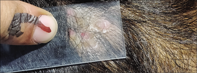
- Apple jelly sign on diascopy in lupus vulgaris
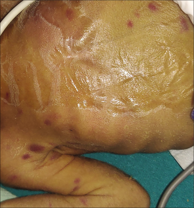
- Blueberry muffin sign in congenital rubella
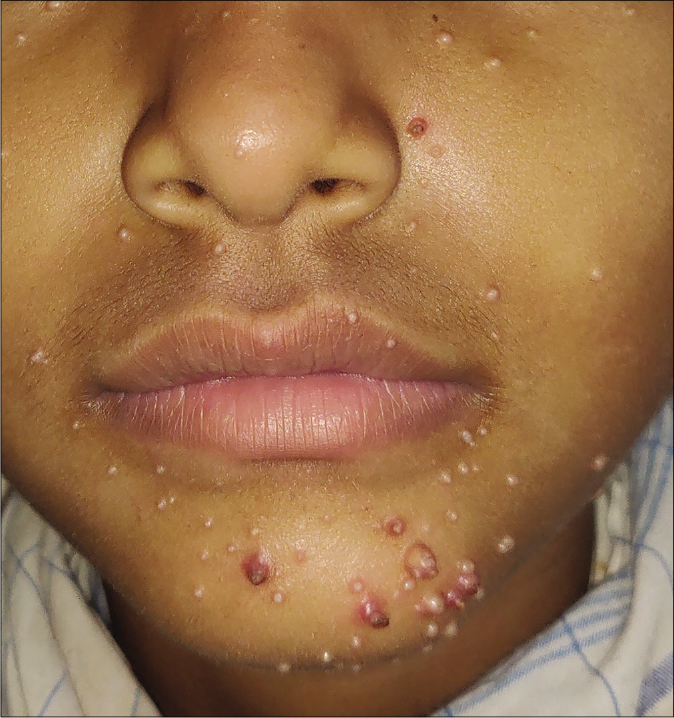
- BOTE sign in molluscum contagiosum
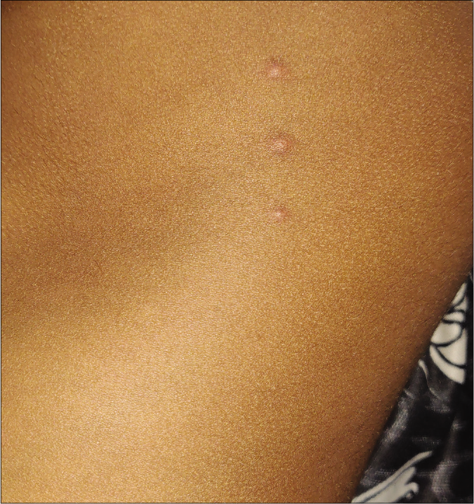
- “Breakfast, lunch and dinner sign” in bed bug infestation
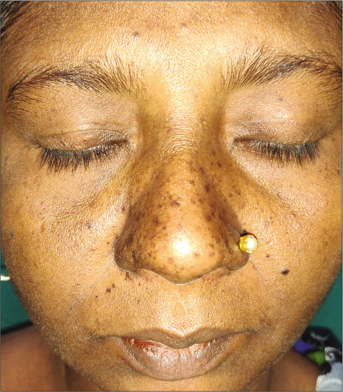
- Centrofacial distribution of pigmentation (chik sign) in a patient of chikungunya
- Dew drop on rose petal appearance seen in varicella
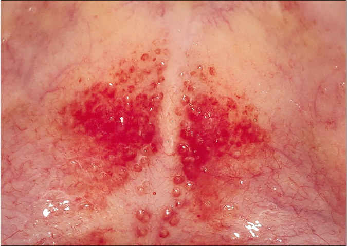
- Erythematous papules with pale center (doughnut sign) in Group A streptococcal pharyngitis
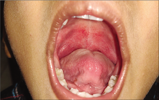
- Forscheimer’s sign in rubella
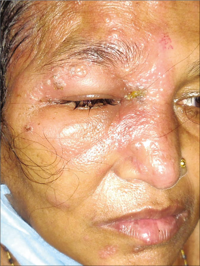
- Involvement of nasal tip in herpes zoster ophthalmicus (Hutchinson’s sign)
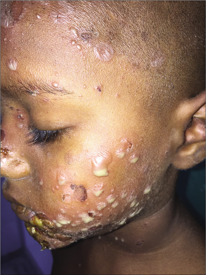
- Hypopyon sign in bullous impetigo (Courtesty- Dr. Abdul Sahid Khan, Department of Dermatology, SCB Medical College, Cuttack, Odisha)

- Papular lesion on palms of patients with secondary syphilis which elicit tenderness on applying pressure (Buschke-Ollendorff sign)
Various signs seen in syphilis and Hansen’s disease are summarized in Tables 1 and 2.
| Sign | Description |
|---|---|
| Buschke-Ollendorff sign | First described by Abraham Buschke and Helene Ollendorff Curth, this sign was named after their observation of slight tenderness on applying pressure with a dull probe on papular lesions of palms in secondary syphilis and is due to cutaneous vasculitis [Figure 11] |
| Barber-pole sign | In congenital syphilis, necrotizing funisitis leads to typical appearance of interspersion of blue and pink areas along with the chalky white appearance in spiral course on umbilical cord, which resembles the barber-pole. |
| Biederman’s sign | A dark red color (instead of the normal pink) of the anterior pillars of the throat, seen in some patients with syphilis |
| Bulldog jaw sign | In congenital syphilis, normal mandible appears relatively longer and bigger due to maxillary hypoplasia |
| Café-au-lait tint sign | Typical brownish-yellow appearance of skin in congenital syphilis due to combined effect of hyperpigmentation, jaundice and anemia |
| Crown of venus sign | These are scaly papules seen along hairline of scalp in secondary syphilis |
| Dory flop sign | When chancre of primary syphilis develops on undersurface of prepuce, then affected part of prepuce does not roll smoothly and rolls out all at once which resembles the movement of dory, a type of fishing boat and is known as dory flop sign |
| Du Bois sign | Shortened little finger, a stigmata of congenital syphilis |
| Elliot sign | Induration of the edge of a syphilitic skin lesion |
| Higoumenaki’s sign | Enlargement of sternal end of (right) clavicle of frequently observed in patients with late congenital syphilis due to periostitis |
| Hutchinson teeth sign: (Screwdriver teeth) |
Abnormal permanent upper central incisors that are peg shaped and notched, usually with obvious thinning and discoloration of enamel in the area of notching; they are widely spaced and shorter than lateral incisors; width of the biting surface is less than that of the gingival margin.It is one of the commonly observed congenital syphilis stigmata |
| Krisowski’s sign | Presence of cicatricial lines radiating from the mouth in congenital syphilis |
| Mulberry molars (moon/Fournier molars) | The biting surface of the first molars becomes dome shaped and has multiple underdeveloped and poorly enameled cusps that occur as a part of congenital syphilis stigmata |
| Natiform skull sign (hot cross bun skull) | Frontoparietal bossing along with prominent suture lines of skull; stigmata due to healed gummatous osteoperiostitis in congenital syphilis |
| Olympian brow sign | Bony prominence of forehead seen in congenital syphilis. Also known as “beetled brow” |
| Omnibus sign | Eyebrow alopecia in secondary syphilis is termed the omnibus sign as it could be seen by a glance at a patient in an omnibus, from the sidewalk |
| Opera-glass nose sign | In congenital syphilis, due to nasal chondritis, the appearance of nose resembles opera glass as the lower part of nose appears to be pushed towards intact upper part |
| Parrot’s nodes | Localized osteoperiostitis of skull vault leads to the formation of round bony swellings in frontoparietal region followed by permanent thickening of bones in late congenital syphilis |
| Sabre shins (Fournier’s sign) | Typical appearance of shins, resembling the sabre sword, in late congenital syphilis and is due to thickening of shaft of the middle third of tibia. |
| Seeping sign | Bleeding from the mucous surfaces and navel, shortly after birth; a sign of syphilis hemorrhagic neonatorum |
| Silex’s sign | A pathognomonic sign of congenital syphilis and is characterized by radial furrows around mouth |
| Split papules | Mucous patches at angle of mouth or corner of nose in early congenital syphilis |
| Virchow’s sign | Tongue with a smooth base seen in patients with congenital syphilis. |
| Sign | Description |
|---|---|
| Benediction sign | Outstretched index and middle fingers with flexion of ring and little fingers due to median nerve involvement in leprosy |
| Elbow sign | Thickened ulnar nerve at the elbow in leprosy |
| Fibula sign | Thickened common peroneal nerve in leprosy |
| Fountain of youth sign | In diffuse lepromatous leprosy, there is smoothening out of patient’s facial wrinkles which restores the youthful appearance |
| Froment sign | Flexion of the distal interphalangeal joint of the thumb when the patient tries to grasp a piece of paper between thumb and index finger; when this is associated with hyperextension of metacarpophalangeal joint of the thumb, it is kown as Jeanne’s sign; indicates weakness of first palmar interossei and adductor pollicis (involvement of deep branch of ulnar nerve) |
| Hertoghe sign (Queen Anne’s sign) |
In lepromatous leprosy, loss of hairs bilaterally in the lateral third of the eyebrows is known as Hertoghe sign or Queen Anne’s sign. This may also be seen in myxedema, follicular mucinosis, atopic dermatitis, trichotillomania, ectodermal dysplasia, discoid lupus erythematosus, alopecia areata, syphilis, ulerythema ophryogenes, systemic sclerosis, HIV and hypothyroidism |
| Wartenberg’s sign | It is one of the earliest signs of ulnar nerve involvement in leprosy and is characterized by constant abduction of small finger; occurs due to paralysis of adductor digiti minimi, which is supplied by the ulnar nerve |
Declaration of patient consent
Patient’s consent not required as there are no patients in this study.
Financial support and sponsorship
Nil.
Conflicts of interest
There are no conflicts of interest.
References
- Blueberry muffin rash at birth due to congenital rubella syndrome. Indian J Paediatr Dermatol. 2013;14:73-5.
- [CrossRef] [Google Scholar]
- Molluscum BOTE sign: A predictor of imminent resolution. Pediatrics. 2013;131:e1650-3.
- [CrossRef] [Google Scholar]
- Linear erythematous papules in a young boy. J Paediatr Child Health. 2020;56:653.
- [CrossRef] [Google Scholar]
- Subconjunctival loaloa with calabar swelling. Indian J Ophthalmol. 2007;55:165-6.
- [CrossRef] [Google Scholar]
- Physical Signs in Medicine and Surgery: An Atlas of Rare, Lost and Forgotten Physical Signs: Includes a Collection of Extraordinary Papers in Medicine, Surgery and the Scientific Method Ocala, FL: Publisher Museum Press Books; 2009.
- [Google Scholar]
- Chikungunya and pigmentation: A tale of two cases. Pigment Int. 2018;5:59.
- [CrossRef] [Google Scholar]
- Pyemotes ventricosus dermatitis, Southeastern France. Emerg Infect Dis. 2008;14:1759-61.
- [CrossRef] [Google Scholar]
- Doughnut lesions over palate in streptococcal pharyngitis. Indian J Pediatr. 2020;87:573.
- [CrossRef] [Google Scholar]
- "Goat eyes": Horizontal rectangular pupils: An unusual clinical presentation of orf. Indian J Dermatol Venereol Leprol. 2015;81:327.
- [CrossRef] [Google Scholar]
- Dermatology eponyms sign lexicon (V) Our Dermatol Online. 2019;10:413-6.
- [CrossRef] [Google Scholar]
- Bilateral groove sign with penoscrotal elephantiasis. Sex Transm Infect. 2002;78:458.
- [CrossRef] [Google Scholar]
- Hanging groin and persistent pruritus in a patient from Burkina Faso. Int J Dermatol. 2007;46:485-6.
- [CrossRef] [Google Scholar]
- Hutchinson's sign as a marker of ocular involvement in HIV-positive patients with herpes zoster ophthalmicus. S Afr Med J. 2010;100:172-4.
- [CrossRef] [Google Scholar]
- The Good Samaritan dispensary of New York city, and the description of Koplik's spots. Arch Pediatr Adolesc Med. 1996;150:535-9.
- [CrossRef] [Google Scholar]
- "Leopard skin" and onchocerciasis. Trans R Soc Trop Med Hyg. 1983;77:881.
- [CrossRef] [Google Scholar]
- Oral manifestations of HIV infection in children. Oral Surg Oral Med Oral Pathol. 1992;73:187-92.
- [CrossRef] [Google Scholar]
- Uvulopalatoglossal junctional ulcers--an early clinical sign of exanthem subitum due to human herpesvirus 6. Med J Malaysia. 1999;54:32-6.
- [Google Scholar]
- Nikolsky's sign in staphylococcal scalded skin syndrome: A new diagnostic clue to the level of epidermal split. Indian J Paediatr Dermatol. 2012;13:51-2.
- [CrossRef] [Google Scholar]
- Bullous skin lesions in Vibrio vulnificus infection. Emerg Med J. 2013;30:863.
- [CrossRef] [Google Scholar]
- Diagnostic value of Pastia's sign in scarlet fever. Cal State J Med. 1912;10:305-6.
- [Google Scholar]
- Eponymous signs in dermatology. Indian Dermatol Online J. 2012;3:159-65.
- [CrossRef] [Google Scholar]
- The Rudolph sign of nasal vestibular furunculosis: Questions raised by this common but under-recognized nasal mucocutaneous disorder. Dermatol Online J. 2012;18:6.
- [CrossRef] [Google Scholar]
- Scaly signs in dermatology. Indian J Dermatol Venereol Leprol. 2006;72:161-4.
- [CrossRef] [Google Scholar]
- Infestation Strongyloides stercoralis in medical personel. N Dermatol Online. 2010;1:32-3.
- [Google Scholar]
- The strawberry tongue: What, how and where? Indian J Dermatol Venereol Leprol. 2018;84:500-5.
- [CrossRef] [Google Scholar]
- Asymmetric periflexuralexanthem in an adult. Clin Exp Dermatol. 2004;29:320-1.
- [CrossRef] [Google Scholar]
- Tasleem's water jet sign a new sign in dermatology. Our Dermatol Online. 2015;6:382-3.
- [CrossRef] [Google Scholar]
- Unusual presentation of cutaneous leishmaniasis. Indian J Dermatol. 2012;57:55-7.
- [CrossRef] [Google Scholar]
- Dengue rash: White islands in a sea of red. Postgrad Med J. 2019;95:676.
- [CrossRef] [Google Scholar]
- Hypothesis: The significance of Winterbottom's sign. J Trop Med Hyg. 1991;94:338-40.
- [Google Scholar]
- Helen Ollendorff Curth and Curth-Macklin syndrome. Open Dermatol J. 2011;5:28-30.
- [CrossRef] [Google Scholar]
- Congenital syphilis In: Venereal Diseases (4th ed). London WC: ELBS and Baillière Tindall; 1980. p. :104-32.
- [Google Scholar]
- Laws and signs of congenital syphilis. J Skin Sex Transm Dis. 2020;2:62-4.
- [CrossRef] [Google Scholar]
- Dermatology eponyms sign lexicon (O) Our Dermatol Online. 2015;6:118-23.
- [CrossRef] [Google Scholar]
- A New Stigma of Hereditary Syphilis. Proceedings of the Medical Society of Athens. :687-99.
- [Google Scholar]
- Wartenberg's sign of ulnar nerve lesion. A contribution to pathophysiology and to the differential diagnosis. Handchir Mikrochir Plast Chir. 2003;35:251-8.
- [Google Scholar]


