Translate this page into:
Cutaneous syndecan-1 expression before and after phototherapy in psoriasis
2 Department of Biochemistry, Faculty of Medicine, Cairo University, Cairo, Egypt
Correspondence Address:
Reham William Doss
Department of Dermatology, Faculty of Medicine, Beni-Suef University, Mohammed Hassan Street, Beni-Suef, 62111
Egypt
| How to cite this article: Doss RW, El-Rifaie AA, Said AN, Rashed LA. Cutaneous syndecan-1 expression before and after phototherapy in psoriasis. Indian J Dermatol Venereol Leprol 2020;86:439-440 |
Sir,
Psoriasis is a multifactorial chronic inflammatory dermatosis, characterized by keratinocyte hyperproliferation, angiogenesis and inflammatory cell infiltrate histologically.
Syndecans are transmembrane proteins that comprise a major family of cell surface heparan sulfate proteoglycans. Syndecan-1, present on the surface of keratinocytes, has the ability to interact with other cell surface receptors and to regulate their actions, controlling cell adhesion, migration and proliferation.[1]
The expression of syndecan is markedly diminished in diseases that exhibit acantholysis, spongiosis or loss of cellular adhesion such as pemphigus vulgaris, bullous pemphigoid and herpes simplex.[1] Its serum levels has also been used as a marker of severity of inflammation in Crohn's disease.[2]
The aim of our study is to investigate the expression of syndecan-1 in the epidermis of lesions of psoriasis and the impact of narrow-band ultraviolet B (NB-UVB) therapy on the level of syndecan-1 in psoriatic epidermis.
The study was done in two steps. Initially, a cross-sectional study was conducted on 20 psoriasis vulgaris patients and 20 age and sex matched healthy volunteers. The study was performed in the dermatology clinic at the faculty of medicine, Beni-Suef University, Egypt. This study protocol was approved by the ethics committee of the Beni-Suef University. The purpose of the study was explained to each patient, and a written consent was taken before data collection. Subjects younger than 15 years or older than 60 years, those suffering from any other acute or chronic illness, malignancies or other inflammatory autoimmune diseases were excluded from the study. Punch skin biopsies (4 mm) were obtained from the back in controls and from a psoriatic plaque in patients and the expression of syndecan-1 was estimated using real time polymerase chaIn reaction (RT-PCR) technique.
This was followed by a therapeutic interventional study. The psoriasis patients were treated with narrow-band UVB (NB-UVB); three sessions per week for a period of 3 months. The starting dose was 50%–70% of the minimal erythema dose (MED) (0.3–0.5 J/cm[2]). The dose was increased at every session till MED was reached after which the dose was kept static. PASI score was estimated for each patient before and after phototherapy. A second punch skin biopsy specimen (4 mm) was taken from the patients after 3 months of phototherapy and the level of syndecan-1 was estimated using RT-PCR technique.
Syndecan-1 was isolated using syndecan-1 extraction kit supplied by mirVana™ PARIS™ Kit, Ambion, USA and then purified and stored at -70°C. Quantification of syndecan-1 was done using RT-PCR technique (TaqMan® syndecan-1 assay).
Statistical analysis was performed with Statistical Package for Scientific Studies (SPSS) 16.0 for Windows (SPSS®, Inc., Chicago, IL, USA). Means and standard deviation values were used to present quantitative data. Mann–Whitney U-test was used to compare quantitative variables. Spearman rank correlation equation for non-normal variables was used to correlate different variables.
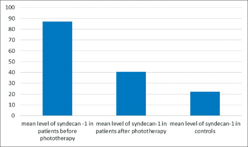
The expression of syndecan-1 in before phototherapy was significantly higher in patients than controls [86.98±23.6 vs. 22.32±4.4 pg/mg respectively; P< 0.001] [Figure - 1].
 |
| Figure 1: Comparison between mean level of expression of syndecan-1 in patients (before and after phototherapy) and controls |
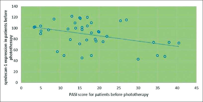
A significant negative correlation was found between the syndecan-1 expression and the PASI score (r =-0.469, and P value = 0.037) [Figure - 2].
 |
| Figure 2: Correlation between the expression of syndecan 1 in psoriatic epidermis before phototherapy and psoriasis areas and severity index |
As previously demonstrated by Alexopoulou et al.[3] and Tomas et al.,[4] syndecan-1 is expressed by proliferating and migrating keratinocytes adjacent to a wound and by psoriatic epidermal cells. The high levels of syndecan-1 expression in psoriasis might be due to keratinocyte proliferation which is the hallmark in psoriasis and as a result of this, high expression inflammatory cells are recruited into psoriatic lesions.
PASI score was calculated for each patient before phototherapy and at the end of the phototherapy sessions for each patient as a marker of severity. Surprisingly The PASI score negatively correlated with the syndecan level meaning that the syndecan level is lower in cases with more severe extensive psoriasis (few cases). In this study, phototherapy helped to reduce the syndecan-1 levels to less than half but the expression still continued to be higher than that of the controls [Figure - 1]. Mean expression in patients before phototherapy 86.98±23.6, after phototherapy 40.56±19.62 vs. 22.32±4.4 pg/mg in controls; P<0.001
The previous studies suggested that the therapeutic effects of phototherapy in psoriasis are conducted through alteration of the cytokine profile and shifting the immune response toward Th2 axis, formation of DNA photoadducts that suppress the accelerated DNA synthesis in psoriatic epidermis inducing apoptosis and DNA damage.[5]
The reduction in syndecan-1 level may be explained by the enhanced matrix metalloproteinase (MMP) production by phototherapy; MMPs are zinc-containing endopeptidases secreted by keratinocytes in response to multiple stimuli such as UV. They are responsible for degrading extracellular matrix proteoglycans including syndecan-1. Another explanation is that NB-UVB induces control of keratinocytes proliferation, and subsequently the reduction in the expression of syndecan-1 on the surface of the basal and suprabasal keratinocytes.
We were unable to find previous studies that investigated the effect of NBUVB on the expression of syndecan-1.
Our study was limited by the small sample size and the lack of prior sample size calculation.
The expression of syndecan 1 could be used as a biomarker for the response of patients of psoriasis to narrow band UVB.
Declaration of patient consent
The authors certify that they have obtained all appropriate patient consent forms. In the form, the patients have given their consent for their images and other clinical information to be reported in the journal. The patient understands that name and initials will not be published and due efforts will be made to conceal identity, but anonymity cannot be guaranteed.
Financial support and sponsorship
Nil.
Conflicts of interest
There are no conflicts of interest.
| 1. |
Bayer-Garner I, Dilday B, Sanderson R, Smoller B. Acantholysis and spongiosis are associated with loss of syndecan-1 expression. J Cutan Pathol 2001;28:135-9.
[Google Scholar]
|
| 2. |
Çekiç C, Kırcı A, Vatansever S, Aslan F, Yılmaz HE, Alper E, et al. Serum Syndecan-1 Levels and Its Relationship to Disease Activity in Patients with Crohn's Disease. Gastroenterol Res Pract 2015;2015:850351.
[Google Scholar]
|
| 3. |
Alexopoulou AN, Multhaupt HA, Couchman JR. Syndecans in wound healing, inflammation and vascular biology. Int J Biochem Cell Biol 2007;39:505-28.
[Google Scholar]
|
| 4. |
Tomas D, Vucić M, Situm M, Kruslin B. The expression of syndecan-1 in psoriatic epidermis. Arch Dermatol Res 2008;300:393-5.
[Google Scholar]
|
| 5. |
Keaney TC, Kirsner RS. New insights into the mechanism of narrow-band UVB therapy for psoriasis. J Invest Dermatol 2010;130:2534.
[Google Scholar]
|
Fulltext Views
2,318
PDF downloads
1,140


