Translate this page into:
Darier's disease following radiotherapy for carcinoma of cervix
2 Johannesburg Hospital, Johannesburg, South Africa
3 Department of Dermatology, Seth G. S. Medical College and KEM Hospital, Mumbai, India
Correspondence Address:
Supriya Chopra
#110, Old building, Tata Hospital, Parel, Mumbai - 400012
India
| How to cite this article: Chopra S, Sharma V, Nischal K C, Khopkar U, Baisane C, Amare (Kadam) P. Darier's disease following radiotherapy for carcinoma of cervix. Indian J Dermatol Venereol Leprol 2004;70:300-303 |
Abstract
Darier-White disease is due to a defect in the ATP2A2 gene encoding the sarcoplasmic/endoplasmic reticulum Ca2+ ATPase (SERCA2b). We report a case of carcinoma cervix in whom Darier's disease manifested after the initiation of radiation therapy. Conventional cytogenetics on peripheral blood revealed non-clonal constitutional autosomal and X chromosome abnormalities suggesting radiation induced gene toxicity. Occurrence of Darier's disease in our case could be due to treatment induced sustained differentiation in the Darier's affected skin by an unknown mechanism. Late onset or sporadic Darier's disease is the other possibility. |
 |
 |
 |
 |
 |
 |
 |
Darier-White disease (DWD), also known as keratosis follicularis (OMIM 124200), is an autosomal dominant disorder characterized by warty papules and plaques in seborrheic areas, palmo-plantar pits and distinctive nail abnormalities. Onset is usually before the third decade with about 95% penetrance and variable expression.[1],[2] We describe here a case of carcinoma of cervix with late onset Darier′s disease.
CASE REPORT
A 46-year-old married woman was seen in the outpatient department of Tata Memorial Hospital. She presented with complaints of bleeding per vaginum for three months. There was no family history of malignancy or any hereditary cutaneous disorder. After detailed clinical, pathological and radiological evaluation she was diagnosed as a case of adenocarcinoma of cervix stage IV-B (in view of metastasis to the supraclavicular nodes). On detailed systemic evaluation there were no other signs of distant metastasis.
She was started on neo-adjuvant chemotherapy and received two cycles of cyclophosphamide and cisplatin followed by concomitant chemotherapy and radiotherapy. The planned dose of radiation was 50 Gy/25 fractions over 5 weeks to the pelvis along with weekly administration of cisplatin. On day 10 of radiation therapy, she developed asymptomatic multiple hyperpigmented keratotic papules associated with itching on the scalp, scapular region, lower abdomen and groin. This was associated with marked mood variations characterized by extreme phases of elation and depression.
Since the rash did not subside, the patient was referred to the Dermatology Department at KEM Hospital after 3 months of radiation. The keratotic papules involved the face, retroauricular region [Figure - 1], upper back and chest accompanied with tiny hypopigmented macules on back and chest. Palmar pits and V-shaped nicking of a few finger nails were observed. The mucosa was uninvolved.
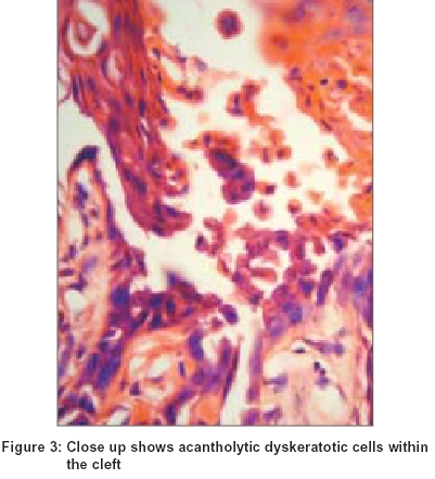
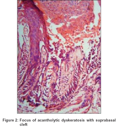
Biopsy from a papule revealed focal epidermal hyperplasia with marked hyperkeratosis, and a few acantholytic cells within suprabasal clefts [Figure - 2]. Scattered dyskeratotic cells (corps ronds) were seen in the stratum malpighii and large parakeratotic dyskeratotic cells with elongated nuclei were seen in the stratum granulosum (grains) [Figure - 3]. The classic clinical and histopathological features established the diagnosis of Darier′s disease even in the absence of a positive family history or prior history of similar lesions.
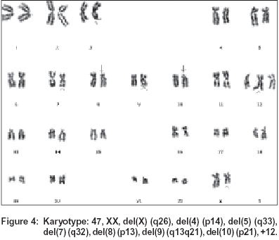
Cytogenetic studies were carried out in phytohemagglutinin (PHA)-M stimulated peripheral blood lymphocytes. Karyotype analysis and aberrations were defined according to ISCN (1995).[3] Of the metaphase plates karyotyped, 60% showed normal diploid karyotype whereas 40% cells had an abnormal karyotypic pattern which included cells with pseudodiploidy, hypodiploidy and hyperdiploidy [Figure - 4]. The most common abnormality was aberration of sex chromosome X, either del (x) (q26) or loss of X.
DISCUSSION
Darier′s disease is caused by the disruption of desmosomal junction, which allows entry of adhesion proteins into the acantholytic cells.[3],[4] The typical histopathological features in Darier′s disease include focal areas of suprabasal clefting with dyskeratotic acantholytic cells (corps ronds and grains) in the epidermis. Electron microscopy reveals loss of desmosomal attachments, perinuclear aggregations of keratin filaments and cytoplasmic vacuolization. These observations suggest that molecules which mediate adhesion between keratinocytes, such as the desmosomal cadherins[5] (upregulation of P-cadherins and down regulation of E-cadherins), desmosomal plaque proteins (desmoplakin, plakoglobin), metalloproteins and their inhibitors or intermediate filament proteins might be involved in the loss of cell-cell adhesion in the epidermis.[6] Alternatively, the defect may lie in a molecule that regulates expression, recruitment, sorting or association of adhesion molecules.
Molecular genetic studies have shown mutation in ATP2A2 at locus 12q23-24.1 as the causative factor.[1],[2] Around 40 different variants of mutant ATP2A2 have been reported. The majority of mutations are nonsense mutations followed by missense mutations.[7] Conventional cytogenetics in our patient revealed common X-chromosomal abnormalities along with abnormalities of other autosomes. Phenotypically the patient was a normal female. The frequency of cells with X-chromosome abnormality was 30% indicating a low degree of constitutional X-chromosome mosaicism which may not affect the phenotype. These constitutional autosomal abnormalities were non-clonal, indicating that they may not be inborn constitutional aneusomies, but could be due to result of radiation related gene toxicity.
Orihuela, et al[8] have described a case of HPV type 16 associated squamous cell carcinoma of the scrotum in a patient with Darier′s disease. However, no direct correlation has been documented between Darier′s disease and HPV 16 associated carcinoma. Vazquez, et al have reported a case of vulvar squamous cell carcinoma arising in localized Darier′s disease.[9]
A case of bronchial carcinoma with preexisting Darier′s disease had exacerbation of lesions within the irradiated area followed by complete clearance after radiation. The authors suggested that radiation induced sustained differentiation of the skin due to an unknown mechanism and speculated on the therapeutic potential of radiation in Darier′s disease.[10]
Darier′s disease is an autosomal dominant condition with variable penetrance. Its occurrence in this patient could be attributed to radiation induced exacerbation of preexisting genetic cohesion defects leading to uncovering of "subclinical" Darier′s disease. The other possibility is that of a late onset sporadic Darier′s disease. A third less probable option is that this late onset or sporadic Darier′s disease may be a cutaneous manifestation of internal malignancy.
| 1. |
Dhitavat J, Dode L, Leslie N. Mutations in the sarcoplasmic/endoplasmic reticulum Ca2+ ATPase isoform cause Darier's disease. J Invest Dermatol 2003;121:486-9.
[Google Scholar]
|
| 2. |
Lowell A Goldsmith, Howard P Baden. Darier- White disease (Keratosis follicularis) and acrokeratosis verruciformis. In: Freedberg IM, Eisen AZ, Wolff K, Austen KF, Goldsmith LA, Katz SI, et al, editors. Dermatology in General Medicine. 5th Ed. New York: McGraw-Hill; 1999. p. 614-7.
[Google Scholar]
|
| 3. |
ISCN (1995). International System for human cytogenetic nomenclature. Mitelman F, editor. Basel: S. Karger, 1995.
[Google Scholar]
|
| 4. |
Hashimoto K, Fujiwara K, Harada M, Setoyama M, Eto H. Junctional proteins of keratinocytes in Grover's disease, Hailey-Hailey's disease and Darier's disease. J Dermatol 1995;22:159-70.
[Google Scholar]
|
| 5. |
Hakuno M, Akiyama M, Shimizu H, Wheelock MJ, Nishikawa T. Upregulation of P-cadherin expression in the lesional skin of pemphigus, Hailey-Hailey disease and Darier's disease. J Cutan Pathol 2001;28:277-81.
[Google Scholar]
|
| 6. |
Burge SM, Schomberg KH. Adhesion molecules and related proteins in Darier's disease and Hailey-Hailey disease. Br J Dermatol 1992;127:335-43.
[Google Scholar]
|
| 7. |
Sakuntabhai A, Ruiz-Perez V, Carter S, Jacobsen N, Burge S, Monk S, et al Mutations in ATP2A2 pump encoding a Ca2+ pump, causes Darier's disease. Nat Genet 1999;21:271-7.
[Google Scholar]
|
| 8. |
Orihuela E, Tyring SK, Pow-Sang M, Dozier S, Cirelli R, Arany I, et al. Development of human papillomavirus type 16 associated squamous cell carcinoma of the scrotum in a patient with Darier's disease treated with systemic isotretinoin. J Urol 1995;153:1940-3.
[Google Scholar]
|
| 9. |
Vazaquez J, Morales C, Gonzalez LO, Lamelas ML, Ribas A. Vulval squamous cell carcinoma arising in localized Darier's disease. Euro J Obstet Gynecol Reprod Biol 2002;102:206-8.
[Google Scholar]
|
| 10. |
MacManus MP, Cavalleri G, Ball DL, Beasley M, Rotstein H, McKay MJ. Exacerbation then clearance of mutation proven Darier's disease of the skin after radiotherapy for bronchial cancer:a case of radiation induced epidermal differentiation. Radiation Research 2001;156:724-30.
[Google Scholar]
|
Fulltext Views
2,500
PDF downloads
1,513


