Translate this page into:
Darier's sign
Correspondence Address:
Amar Surjushe
Department of Dermatology, Venereology, and Leprology, Grant Medical College and Sir JJ Groups of Hospitals, Mumbai - 400008
India
| How to cite this article: Surjushe A, Jindal S, Gote P, Saple D G. Darier's sign. Indian J Dermatol Venereol Leprol 2007;73:363-364 |
 |
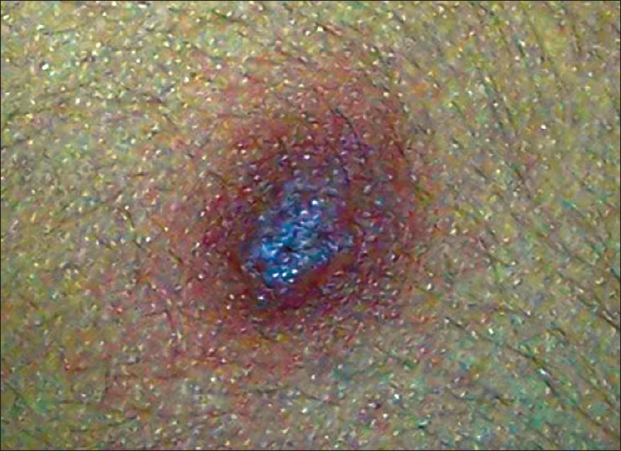
| Figure 1: Darier�s sign |
 |
| Figure 1: Darier�s sign |
Introduction
Darier′s sign refers to the urtication and erythematous halo that are produced in response to rubbing or scratching of lesions of cutaneous mastocytosis.
Darier′s sign is named after the French dermatologist Ferdinand-Jean Darier who first described it. The first description of mastocytosis was made by Nettleship and Tay [1] in 1869, and in 1878, Sangster coined the term urticaria pigmentosa. [2]
Method of Elicitation
In classical Darier′s sign, gentle rubbing or stroking of the lesions, is followed by local itching, erythema and weal formation within 2-5 min. This may persist from 30min to several hours. In young children, there may be vesiculation in the stroked lesion. Although classically positive in lesional skin, this sign may even be demonstrated on clinically normal skin in patients of mastocytosis.
In pseudoxanthomatous mastocytosis, a variant of diffuse cutaneous mastocytosis, there will be only erythema without urtication on rubbing as against a classic Darier′s sign. [3]
Pathophysiology
In cutaneous mastocytosis, there is an increased number of functionally normal mast cells in the dermis. When the skin is stroked, there is degranulation of the mast cells with the release of inflammatory granules that contain histamine, slow-releasing substance of anaphylaxis (SRSA), eosinophil chemotactic activating factor (ECAF) and heparin [Figure - 1]. It is the histamine that is responsible for the response.
It is also suggested that besides exocytosis of single or multiple granules, there is another pattern of degranulation when mast cells rupture. This happens following physical stimuli such as stroking. A gentle action gives rise to exocytosis, while a strong action leads to rupture of the cells. [4]
Exact pathophysiology of Darier′s sign in lymphoma and leukemias, is not known. One hypothesis suggested that mast cell proliferation may be related to lymphokine release, thereby explaining their close relation with lymphocytes. [5] Another hypothesis suggested that laminin increases mast cell attachment to the lymphocytes. [6] In juvenile xanthogranuloma, the histiocytes of the lesions undergo macrophagic differentiation to dermal dendrocytes, and the distribution of mast cells in the skin coincides with that of dermal dendritic cells. [7]
Conditions Associated With Darier′s Sign
- Cutaneous mastocytosis: In urticaria pigmentosa, the most frequent clinical form of cutaneous mastocytosis, Darier′s sign was present in 94% of cases. [8]
- Leukemia cutis: Leukemia cutis occurs in 25-30% of infants with congenital leukemia and is frequently associated with acute myeloid leukemia than with acute lymphoblastic leukemia. Urticaria-pigmentosa-like lesions have been reported in acute lymphoblastic leukaemia. [9],[10]
- Juvenile xanthogranuloma: It is the most common form of non-Langerhans cell histiocytosis. Nagayo et al. reported Darier′s sign in this disorder. [7]
- Histiocytosis X : Foucar et al. described positive Darier′s sign in a patient with ′mast cell rich variant′ of histiocytosis X. [11]
- Lymphoma: On rare instancs, Darier′s sign has been reported in cutaneous large T-cell lymphoma and in non-Hodgkin′s lymphoma. [12],[13]
Differential Diagnosis
- Pseudo-Darier′s sign : It is a transient piloerection and elevation or increased induration of a lesion induced by rubbing and is observed in congenital smooth muscle hamartomas. A positive pseudo-Darier′s sign can be helpful in clinically distinguishing congenital smooth muscle hamartoma from congenital hairy nevus. [14]
- Dermatographism ("skin writing"): It is a form of physical urticaria that consists of local erythema due to capillary vasodilatation, followed by edema and a surrounding flare due to axon reflex induced dilation of arterioles, which is observed after the firm stroking of skin. The cause of this phenomenon is thought to be hypersensitivity of the mast cells rather than an increase in the mast cells, as observed in mastocytosis. [15]
Significance
Darier′s sign is pathognomic of cutaneous mastocytosis although some patients may have little or no wealing or itching even when the skin reveals a dense population of mast cells, particularly in those with a long history of the disorder. However, Darier′s sign is not 100% specific for mastocytosis since it has been described, albeit rarely, in juvenile xanthogranulomas and acute lymphoblastic leukemia. [15]
Acknowledgement
We acknowledge Department of Dermatology of Rajiv Gandhi Medical College, Mumbai for providing us the photograph of Darier′s sign.
| 1. |
Nettleship E, Tay W. Rare forms of urticaria. BMJ 1869;2:323-30.
[Google Scholar]
|
| 2. |
Sangster A. An anomalous mottled rash, accompanied by pruritus, facticious urticaria and pigmentation, 'urticaria pigmentosa (?). Trans Clin Soc London 1878;11:161-3.
[Google Scholar]
|
| 3. |
Beer WW, Emslie ES. Diffuse cutaneous mastocytosis. Br J Dermatol 1968;80:841-3.
[Google Scholar]
|
| 4. |
Zhu KJ, Zhu TC, Shi YH, Ge ZH. A histopathological and electron microscopical observation of urticaria pigmentosa. Indian J Dermatol Venereol Leprol 1990;56:152-5.
[Google Scholar]
|
| 5. |
Prokocimer M, Polliack A. Increased bone marrow mast cells in preleukemic syndromes, acute leukaemia and lymphoproliferative disorders. Am J Clin Pathol 1981;75:34-8.
[Google Scholar]
|
| 6. |
Thompson HL, Burbelo PD, Segui-Real B, Yamada Y, Metcalfe DD. Laminin promotes mast cell attachment. J Immunol 1989;143:2323-7.
[Google Scholar]
|
| 7. |
Nagayo K, Sakai M, Mizuno N. Juvenile xanthogranuloma with Darier's sign. J Dermatol 1983;10:283-5.
[Google Scholar]
|
| 8. |
Kiszewski AE, Durαn-Mckinster C, Orozco-Covarrubias L, Gutiιrrez-Castrell�n P, Ruiz-Maldonado R. Cutaneous mastocytosis in children: A clinical analysis of 71 cases. J Eur Acad Dermatol Venereol 2004;18:285-90.
[Google Scholar]
|
| 9. |
Yen A, Sanchez R, Oblender M, Raimer S. Leukemia cutis: Darier's sign in a neonate with acute lymphoblastic leukemia. J Am Acad Dermatol 1996;34:375-8.
[Google Scholar]
|
| 10. |
Raj S, Khopkar U, Wadhwa SL, Kapasi A. Urticaria-pigmentosa-like lesions in acute lymphoblastic leukaemia (2 cases). Dermatology 1993;186:226-8.
[Google Scholar]
|
| 11. |
Foucar E, Piette WW, Tse DT, Goeken J, Olmstead AD. Urticating histiocytosis: A mast cell rich variant of histocytosis X. J Am Acad Dermatol 1986;14:867-73.
[Google Scholar]
|
| 12. |
Ollivaud L, Cosnes A, Wechsler J, Gaulard P, Bagot M, Haioun C, et al . Darier's sign in cutaneous large T-cell lymphoma. J Am Acad Dermatol 1996;34:506-7.
[Google Scholar]
|
| 13. |
Lewis FM, Colver GB, Slater DN, Darier's sign associated with non-Hodgkin's lymphoma. Br J Dermatol 1994;130:126-7
[Google Scholar]
|
| 14. |
Goldman MP, Kaplan RP, Heng MC. Congenital smooth muscle hamartoma. Int J Dermatol 1987;26:448-52.
[Google Scholar]
|
| 15. |
Grattan CEH, Black AK. Urticaria and mastocytosis. In: Burns T, Breathnach S, Cox N, Griffiths G, editors. Rook's Textbook of Dermatology. 7 th ed. Blackwell publishing; 2004. p. 47.1- 47.37."
th ed. Blackwell publishing; 2004. p. 47.1- 47.37."'>[Google Scholar]
|
Fulltext Views
23,630
PDF downloads
4,710


