Translate this page into:
Deep morphea in a child after pneumococcal vaccination
Correspondence Address:
Elena del Alcazar Viladomiu
Department of Dermaology, Hospital Universitario Donostia Paseo Dr. Begiristain 107-116, Edificio Amara 2� Planta, 20014 Donostia-San Sebasti�n, Gipuzkoa
Spain
| How to cite this article: Viladomiu Ed, Valls AT, Zabaleta BA, Moreno AJ, P�rez NO. Deep morphea in a child after pneumococcal vaccination. Indian J Dermatol Venereol Leprol 2014;80:259-260 |
Sir,
Although the etiology of morphoea is unknown without a remarkable different trigger factors, including intramuscular vaccine injections have been reported. We have found only 13 published cases of post-vaccination morphea. [1],[2],[3],[4],[5] We report a case of morphea in a child after receiving the second dose of pneumococcal vaccination.
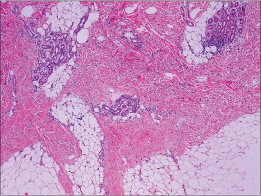
A 12-month-old Colombian boy, with an unremarkable medical history, was sent to our department for an indurated plaque on his left thigh. His parents reported that the induration had appeared at around 6 months of age, 20 days after receiving the second dose of intramuscular pneumococcal vaccination. Initially the lesion grew, stabilizing 2 months later. Cutaneous examination showed an indurated plaque that measured 12 cm × 6 cm in diameter, with poorly defined edges [Figure - 1]. The overlying epidermis was normal. The mobility of the knee and hip was preserved. Physical examination was otherwise normal. A skin biopsy showed increased dermal thickness and homogenization with eosinophilic and thick collagen bundles without fibrosis accompanied by a mild perivascular and peri-adnexal lymphocytic infiltrate [Figure - 2]. Laboratory studies including blood cell count, serum chemistry, autoantibodies (antinuclear antibodies and anti-SCl70) and Borrelia burgdoferi serology were normal or negative. Clinical and pathological features were consistent with the diagnosis of deep morphea. Treatment with local clobetasol for 4 weeks was ineffective and was stopped. At the moment, the plaque remains stable and the growth of both limbs appears to be identical.
 |
| Figure 1: Deep induration of 12 × 6 cm with typical orange-peel appearance of the left thigh |
 |
| Figure 2: Perivascular and peri-adnexal lymphocitic infi ltrate, with dermal homogenization, and eosinophilic and thick collagen bundles infi ltrating the subcutaneous adipose tissue (H and E, ×40) |
Post-vaccination morphea is rarely reported in the medical literature; we found only 13 reported cases. The vaccines implicated in previous reports are: Bacillus Calmette-Guérin (BCG) (1 case), diphtheria-tetanus-pertussis (DTP) (3 cases), tetanus (2 cases), measles, mumps and rubella (MMR) (1 case) and hepatitis B vaccine (5 cases). [1],[2],[3],[4] In an article in 2011, Kumar and Noronha described a new case without an identified vaccine. [5]
In our case, the vaccine implicated was pneumococcal vaccine, a polysaccharide conjugate vaccine that contains extracts from 13 of the most common types of Streptococcus pneumoniae bacteria. Pneumococcal vaccine is usually recommended to be used in babies and young children from 6 weeks to 5 years of age and children normally receive four doses of the vaccine, starting at 6 weeks of age.
After vaccination, different types of morphea are reported, deep morphea (six cases) being the most frequent. [1],[2],[3],[4] It can involve subcutaneous tissue, fascia and muscle and can produce contractures and deformities. Other types of morphea reported are plaque morphea (four cases), [1],[5] generalized morphea (one case) [1] and sclerodermiform reaction (two cases). [1]
The pathogenesis is unknown though Torrelo et al. suggested that vaccines may induce an immune response against specific and non-specific antigens in patients with trigger factors and that the trauma of injection may cause endothelial damage and tissue hypoxia. [1]
Different treatments have been tried including potent topical corticosteroids, systemic corticosteroids and methotrexate but none has proved effective. [1]
Even though no case of progression to generalized morphea or of the development of systemic symptoms has been reported, we consider it important to follow-up the patient in order to assess the symmetric growth of the limbs and detect any appearance of systemic symptoms. [4]
| 1. |
Torrelo A, Suárez J, Colmenero I, Azorín D, Perera A, Zambrano A. Deep morphea after vaccination in two young children. Pediatr Dermatol 2006;23:484-7.
[Google Scholar]
|
| 2. |
Khaled A, Kharfi M, Zaouek A, Rameh S, Zermani R, Fazaa B, et al. Postvaccination morphea profunda in a child. Pediatr Dermatol 2012;29:525-7.
[Google Scholar]
|
| 3. |
Benmously Mlika R, Kenani N, Badri T, Hammami H, Hichri J, Haouet S, et al. Morphea profunda in a young infant after hepatitis B vaccination. J Am Acad Dermatol 2010;63:1111-2.
[Google Scholar]
|
| 4. |
Bukhari I, Al Breiki S. Post vaccination localized morphea. J Chin Clin Med 2009;4:599-600.
[Google Scholar]
|
| 5. |
Kumar P, Noronha P. Post vaccination morphea in a child. J Pediatr Sci 2011;3:2-5.
[Google Scholar]
|
Fulltext Views
4,174
PDF downloads
2,752


