Translate this page into:
Dermoscopic features of a flesh-coloured papule on the tongue
Corresponding author: Dr. Madhusmita Sethy, Department of Pathology, All India Institute of Medical Sciences, Bhubaneswar, Odisha, India. drmaheshmadhu@gmail.com
-
Received: ,
Accepted: ,
How to cite this article: Behera B, Dash S, Mishra JJ, Viswan P, Sethy M. Dermoscopic features of a flesh-coloured papule on the tongue. Indian J Dermatol Venereol Leprol 2023;89:870.
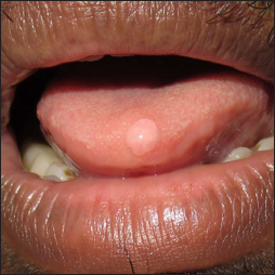
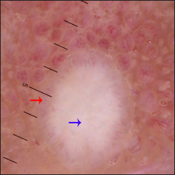
A 30-year-old male had a single asymptomatic, slow-growing lesion on the tip of the tongue for six months. He denied any prior history of trauma or surgical procedure at the site. His medical history was unremarkable. Cutaneous examination showed a solitary well-demarcated flesh-coloured, dome-shaped, smooth, firm, non-tender papule of size 4 mm × 4 mm on the tip of the tongue [Figure 1]. The dermoscopic examination under polarised mode (Heine Delta 20T, ×10 magnification) revealed a central white homogeneous area and a peripheral corona of hairpin vessels [Figure 2]. The differential diagnoses of traumatic fibroma, granular cell tumour, Heck’s disease, and angiofibroma were considered. Pathological examination showed a moderately acanthotic epidermis, a collagenous stroma with numerous dispersed dilated blood vessels and scattered fibroblasts. In addition, inflammatory cell infiltration was absent. The diagnosis of traumatic fibroma was made, and the patient was counselled about the benign nature. The white homogeneous area corresponds to the acanthotic epidermis along with dermal collagen and the hairpin vessels to the dilated papillary dermal blood vessels.

- Solitary flesh-coloured, dome-shaped, smooth papule on the tip of the tongue

- The dermoscopic examination under polarized mode (Heine Delta 20T, ×10) shows a central white homogeneous area (blue arrow) and a peripheral corona of hairpin vessels (red arrow)
Declaration of patient consent
The authors certify that they have obtained all appropriate patient consent.
Financial support and sponsorship
Nil.
Conflict of interest
There are no conflicts of interest.


