
Translate this page into:
Dermoscopic features of cutaneous metastases from breast carcinoma: A report of three Indian patients
-
Received: ,
Accepted: ,
How to cite this article: Gupta V, Bhatia R, Taneja N, Mridha AR. Dermoscopic features of cutaneous metastases from breast carcinoma: A report of three Indian patients. Indian J Dermatol Venereol Leprol 2021;87:273-8.
Sir,
Dermoscopic features of cutaneous metastases from internal malignancies have been documented in only a few reports, predominantly in fair-skinned population.1,2 Here, we describe the dermoscopic features of cutaneous metastases from breast carcinoma in three Indian patients.
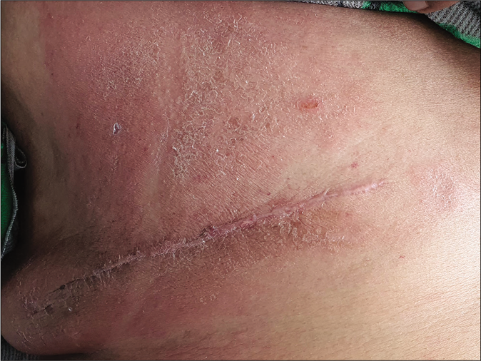
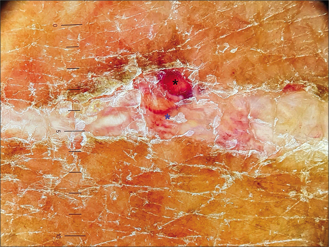
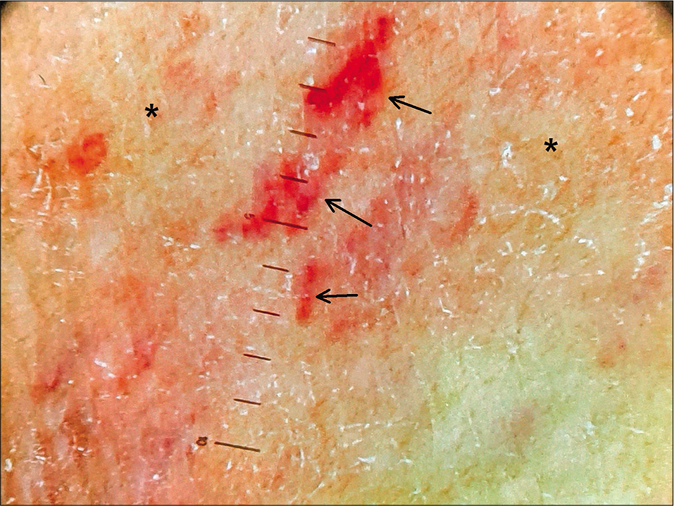
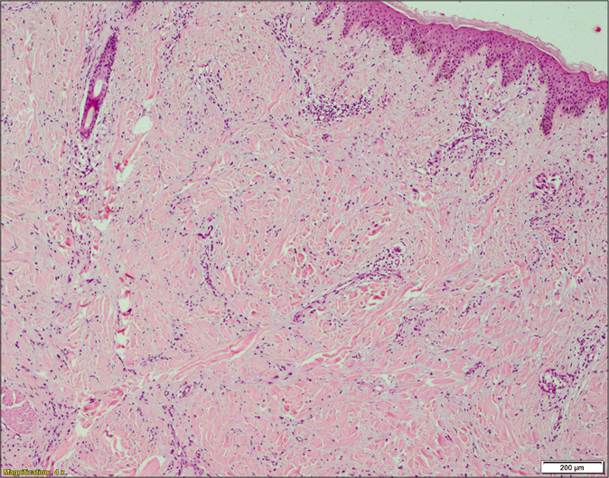
Case 1: A 36-year-old female, with a previous history of left breast carcinoma treated with modified radical mastectomy and adjuvant radiotherapy one year back, presented with skin-coloured translucent to hyperpigmented papules and clear fluid-filled vesicles on the left mammary region and abdomen for the last three months. These skin lesions appeared to be arranged in linear horizontal bands, on a background of ill-defined reddish-brown induration along with focal areas of depigmentation and overlying erosions [Figure 1]. Polarized dermoscopy of the papules and vesicles showed well-defined yellow-white lacunae separated by thin brownish septae, some of which had adjacent smaller red lacunae. Background showed erythematous structureless areas and multiple scattered small round whitish clods. The depigmented lesions showed structureless whitish areas with pinkish hue and mild scaling [Figure 2]. Skin biopsy from the plaque as well as papule showed tumour nests in the dermis. Lymphovascular invasion was noted in the biopsy from papule [Figure 3].

- Patient 1: Skin-coloured to hyperpigmented papules and clear fluid-filled vesicles on the left mammary region and abdomen on a background of ill-defined reddish-brown induration. There are focal areas of depigmentation and overlying erosions
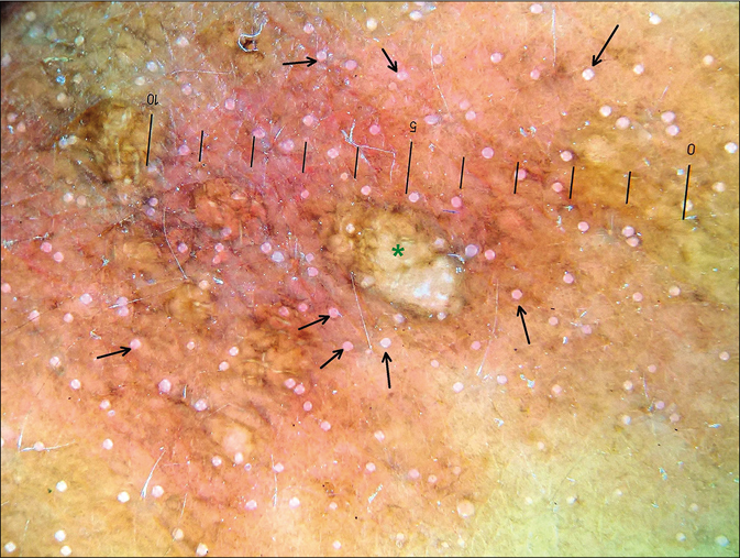
- Patient 1: Dermoscopic examination (polarized, ×16; Heine Delta 20T, Heine Optotechnik, Herrsching, Germany) of a translucent papule on breast showing yellow-white lacunae separated by thin brownish septae (green star), with background erythematous structureless areas and multiple, scattered small round whitish clods (black arrows)
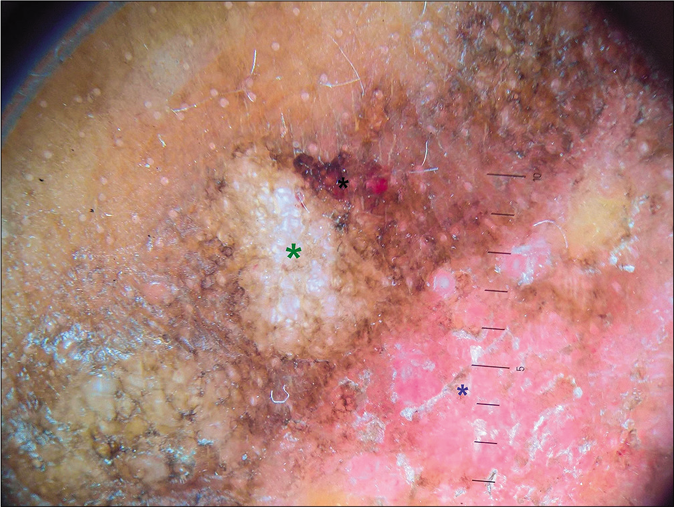
- Patient 1: Dermoscopic examination (polarized, ×16; Heine Delta 20T, Heine Optotechnik, Herrsching, Germany) of a papule on abdomen showing yellow-white lacunae separated by thin brownish septae (green star) with adjacent small red lacunae (black star), structureless whitish areas with pinkish hue and mild scaling (blue star)
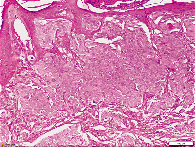
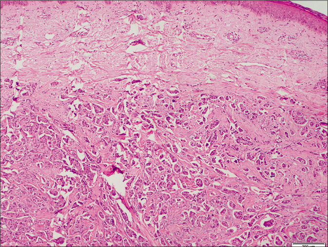
- Patient 1: Skin biopsy shows sheets of tumor cells arranged in groups and clusters involving the papillary dermis. Cells are polygonal with large hyperchromatic nuclei, prominent eosinophilic nucleoli and high nuclear-cytoplasmic ratio. Brisk mitosis are present (H and E, ×100)
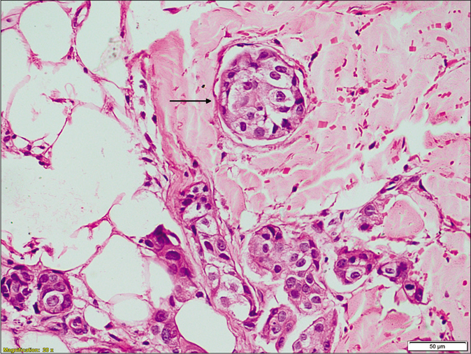
- Tumour emboli within the vascular lumen (black arrow) in deeper dermis (H & E, x200)
Case 2: A 43-year-old female presented to us with ill-defined, erythematous, firm plaques on the right mammary region [Figure 4] for 1 month. She had been diagnosed with bilateral breast carcinoma eight months back, which had been treated with neoadjuvant chemotherapy and bilateral modified radical mastectomy. Polarized dermoscopy showed short, branched linear vessels focally and a background erythematous structureless areas, while the mastectomy scar showed reddish clods and adjacent short curvilinear vessels [Figure 5]. Skin biopsy showed diffuse dermal pattern of metastases from breast carcinoma [Figure 6].
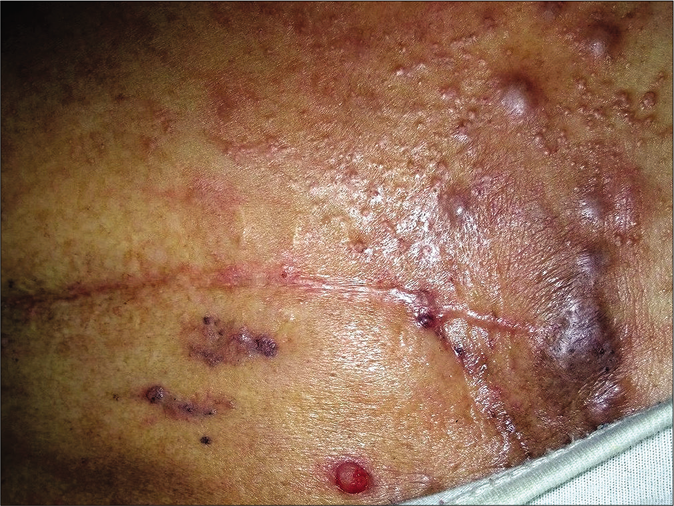
- Patient 2: Ill-defined, erythematous, firm plaques on the right mammary region
- Patient 2: Dermoscopic examination (polarized, ×16; Heine Delta 20T, Heine Optotechnik, Herrsching, Germany) of the skin over mastectomy scar showing reddish clods (black star) and adjacent short curvilinear vessels (blue star)
- Patient 2: Dermoscopic examination (polarized, ×16×; Heine Delta 20T, Heine Optotechnik, Herrsching, Germany) of the ill-defined plaque showing short branched linear vessels focally (black arrows) and background erythematous structureless areas (black star)
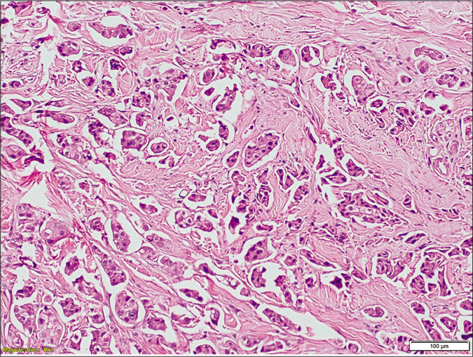
- Patient 2: H and E stained sections show an infiltrate in diffuse pattern in the dermis (×40)
- Higher magnification showing isolated tumor cells with centrally located hyperchromatic nucleolus and moderate amount of eosinophilic cytoplasm (H and E, ×400)
Case 3: A 54-year-old female, with right breast carcinoma operated 18 months back, presented to us with grouped, skin-coloured, firm papules and violaceous papules and nodulo-plaques on the right mammary region, along with pitting edema of the right arm [Figure 7]. Polarized dermoscopy of the violaceous plaque showed asymmetric blue-black ovoid nests of variable size, focal clustered gray dots and pigment network, an erythematous structureless zone with parallel whitish streaks and short branched vessels. Skin coloured papules showed branching linear vessels on a background of structureless erythematous areas [Figure 8]. Skin biopsy confirmed the diagnosis of breast carcinoma cutaneous metastases [Figure 9].
- Patient 3: Grouped skin-coloured firm papules and violaceous papules and nodulo-plaques on the right mammary region
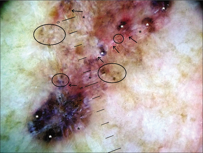
- Patient 3: Dermoscopic examination (polarized, ×16; Heine Delta 20T, Heine Optotechnik, Herrsching, Germany) of the violaceous plaque showing asymmetric blue-black ovoid nests of variable size (white stars), focal clustered gray dots and pigment network (black circles), an erythematous structureless zone with parallel whitish streaks (black stars) and short branched vessels (black arrows)
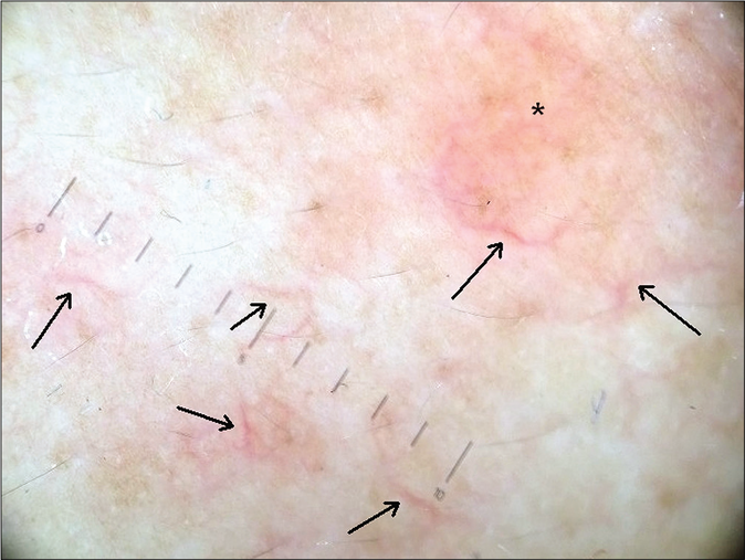
- Patient 3: Dermoscopic examination (polarized, ×16; Heine Delta 20T, Heine Optotechnik, Herrsching, Germany) of the skin coloured papule showing branching linear vessels (black arrows) on a background of structureless erythematous areas (black stars)
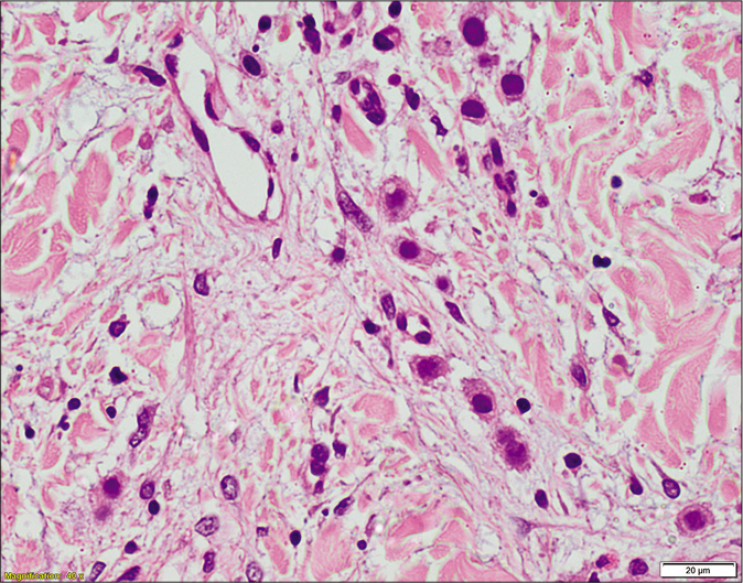
- Patient 3: Tumor involves the primarily deep dermis, and the cells are arranged in small groups (H and E, ×400)
- Higher magnification shows the tumour cells are cuboidal with large hyperchromatic nuclei, prominent eosinophilic nucleoli and high nuclear-cytoplasmic ratio (H and E, ×100)
Dermoscopic features of cutaneous tumours such as basal cell carcinoma, Bowen’s disease and melanoma are well-described; however, there is relative paucity of literature regarding cutaneous metastases from internal organ malignancies. Chernoff et al. reported dermoscopic findings in cutaneous metastases arising from various underlying malignancies in 20 patients, six of whom had breast carcinoma. Of these, 17 lesions were non-pigmented and 3 were pigmented. Dermoscopy of the non-pigmented lesions was characterized by a vascular pattern, most frequently serpentine (linear irregular) vessels (n = 13/17), followed by branched (n = 9/17), dotted (n = 4/17) or comma-shaped vessels (n = 3/17), and homogenous pink areas without discrete vessels (n = 2/17). More than half (59%, n = 10/17) of the lesions showed a polymorphous vascular pattern. The three pigmented lesions, all breast carcinoma metastases, showed melanocytic patterns comprising of pigmented streaks (n = 3), blue– gray globules (n = 2) and a blue-white veil (n = 1).1 Dermoscopy of pigmented cutaneous metastases of breast carcinoma can resemble malignant melanoma or basal cell carcinoma.3,4 Vascular pattern such as linear irregular branched or polymorphic vessels was the predominant finding in two other reports of four cases of non-pigmented breast carcinoma cutaneous metastases, suggesting a role of neo-angiogenesis in their pathogenesis. Other findings included yellow central areas, white lines at periphery and surrounding whitish structureless areas. The peau d’orange appearance showed small umbilicated pits with a tendency to form linear fissure-like structures.2,5
To the best of our knowledge, dermoscopic features of cutaneous metastases have not been described in dark-skinned population. We report the dermoscopic findings of breast carcinoma cutaneous metastases in three Indian patients with Fitzpatrick skin phototype IV or V. The dermoscopic features noted in our patients are similar to the descriptions in previous reports: vascular patterns (linear branched vessels, short curvilinear vessels, red clods, erythematous structureless areas) in skin-coloured or erythematous lesions, and melanoma-like features (asymmetric blue-black ovoid nests, focal gray dots and pigment network, whitish streaks, telangiectasias) in a pigmented lesion.6,7 We also noted yellow-white lacunae and smaller red lacunae separated by thin septae, the dermoscopic features described for lymphangiectasias, probably correlating with the lympho-vascular invasion by tumour cells; these features have not been described for breast carcinoma cutaneous metastases before to our knowledge [Table 1].8,9
| Patient number | Histological type of breast carcinoma | Clinical morphology of cutaneous breast cancer metastases | Histological pattern of metastases | Dermoscopy findings |
|---|---|---|---|---|
| 1 | Invasive ductal carcinoma, no special type | Erythematous indurated plaques | Sheets of tumour cells in the upper dermis | Multiple scattered small round whitish clods, erythematous structureless areas |
| Skin coloured translucent papules and vesicles | Sheets of tumour cells in the upper dermis, along with lymphovascular invasion | Well-defined yellow-white lacunae separated by septae, adjacent smaller red lacunae | ||
| 2 | Invasive ductal carcinoma, no special type | Ill-defined erythematous plaques | Diffuse interstitial infiltrate in dermis | Short-branched linear vessels focally, background erythematous structureless areas |
| Mastectomy scar | Biopsy not done | Reddish clods, short curvilinear vessels | ||
| 3 | Invasive ductal carcinoma, no special type | Skin coloured papules | Tumour islands in deep dermis | Branching linear vessels on a background of structureless erythematous areas |
| Violaceous papules and plaques | Biopsy not done | Asymmetric, blue-black ovoid nests, focal clustered gray dots and pigment network, erythematous structureless zone with parallel whitish streaks and short branched vessels |
The dermoscopic findings of breast carcinoma cutaneous metastases appear to be relatively non-specific, and can resemble benign as well as malignant conditions. However, some dermoscopic features such as linear branched vessels, and features resembling those of melanoma or lymphangiectasias can raise the suspicion of a metastatic deposit in the appropriate setting.
Declaration of patient consent
The authors certify that they have obtained all appropriate patient consent.
Financial support and sponsorship
Nil.
Conflicts of interest
There are no conflicts of interest.
References
- Dermoscopic findings in cutaneous metastases. JAMA Dermatol. 2014;150:429-33.
- [CrossRef] [PubMed] [Google Scholar]
- Dermoscopy of skin metastases from breast cancer and of the orange peel type ("peau d'orange"): A report of two cases. Int J Dermatol. 2015;54:343-6.
- [CrossRef] [PubMed] [Google Scholar]
- Pigmented skin metastasis of breast cancer showing dermoscopic features of malignant melanoma. J Eur Acad Dermatol Venereol. 2015;29:1034-6.
- [CrossRef] [PubMed] [Google Scholar]
- Dermoscopy of pigmented invasive ductal carcinoma mimicking basal cell carcinoma. Australas J Dermatol. 2017;58:326-7.
- [CrossRef] [PubMed] [Google Scholar]
- Dermoscopy of skin metastases from breast cancer: Two case reports. J Med Case Rep. 2018;12:273.
- [CrossRef] [PubMed] [Google Scholar]
- Frequency and morphologic characteristics of invasive melanomas lacking specific surface microscopic features. Arch Dermatol. 1996;132:1178-82.
- [CrossRef] [PubMed] [Google Scholar]
- The significance of crystalline/chrysalis structures in the diagnosis of melanocytic and nonmelanocytic lesions. J Am Acad Dermatol. 2012;67:194.e1-8.
- [CrossRef] [PubMed] [Google Scholar]
- Dermoscopy of lymphangioma circumscriptum: A morphological study of 45 cases. Australas J Dermatol. 2018;59:e189-93.
- [CrossRef] [PubMed] [Google Scholar]
- Localized acquired lymphangiectasias after breast surgery: Enhanced non invasive diagnosis using dermoscopy and reflectance confocal microscopy. Skin Res Technol. 2020;26:205-8.
- [CrossRef] [PubMed] [Google Scholar]


