
Translate this page into:
Dermoscopic features of primary cutaneous extranodal natural killer (NK)/T-cell lymphoma
Corresponding author: Dr. Xiaopo Wang, Department of Pathology, Hospital for Skin Disease, Chinese Academy of Medical Sciences and Peking Union Medical College, Nanjing, China. 13770757675@163.com
-
Received: ,
Accepted: ,
How to cite this article: Wang X. Dermoscopic features of primary cutaneous extranodal natural killer (NK)/T-cell lymphoma. Indian J Dermatol Venereol Leprol. 2025;91:S46-S49. doi: 10.25259/IJDVL_1222_2023
Dear Editor,
The extranodal natural killer (NK)/T-cell lymphoma (ENKTL) derived from NK cells or cytotoxic T cells is a highly aggressive malignancy related to Epstein-Barr virus infection. ENKTL is categorised into two groups: nasal ENKTL which affects the upper aerodigestive tract, and extra-nasal ENKTL affecting the skin. Cutaneous ENKTL can be divided into two distinct subsets: primary cutaneous, which initially presents in the skin, or nasal ENKTL with secondary spread to the skin.1 Dermoscopy may be useful in revealing the possibility of cutaneous lymphoproliferative disorders,2 thereby prompting an early skin biopsy. Herein, we describe the dermoscopic features of primary cutaneous ENKTL, which have not been described previously.
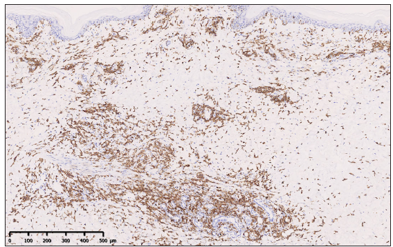
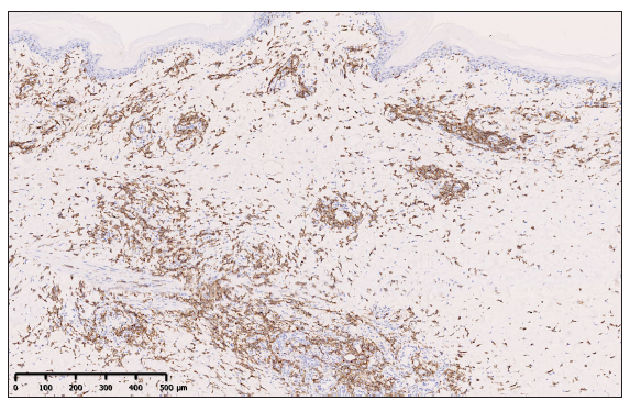
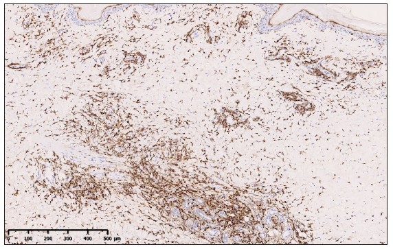
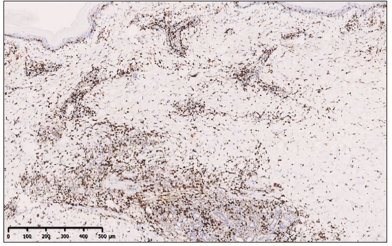
A 64-year-old man presented with a 3-month history of multiple, erythematous skin lesions on his face and limbs. He had been experiencing fever, fatigue, and weight loss for the past month. On physical examination, numerous, indurated erythematous nodules, and plaques were found on the face and extremities [Figures 1a and 1b]. The lesions were slightly painful. The patient was initially diagnosed as acute febrile neutrophilic dermatosis. All laboratory investigations were within normal limits, including blood cell counts and biochemical tests. Dermoscopic features of the eyebrow showed white areas surrounding follicular plugging, with a few short linear vessels at the periphery of the follicles. Scales and a few broken hairs were seen along with pinkish areas [Figure 1c]. On the right lower limb, dermoscopy revealed scales, along with distortion of the pigment network, and dotted or fine linear vessels over an orange-yellowish background [Figure 1d]. In addition, dermoscopy observed a homogeneous dull‐red background, dotted and hairpin vessels with scales on the left lower limb [Figure 1e]. Histopathologic examination of the left lower limb revealed a dermal and subcutaneous infiltration of atypical lymphoid cells, with angiocentricity [Figures 2a and 2b]. Immunohistochemical studies showed that atypical lymphoid cells were positive for CD3, CD8, CD56, and granzyme B [Figures 3a-d], but negative for CD4, CD20, PAX-5, and CD30. The ki-67 was 80% and EBV was demonstrated by in situ hybridisation [Figures 3e and 3f]. Further examination, including computed tomography of other systems, bone marrow biopsy and aspirate, did not uncover any systemic involvement. Following the above findings, he was diagnosed with a primary cutaneous ENKTL.
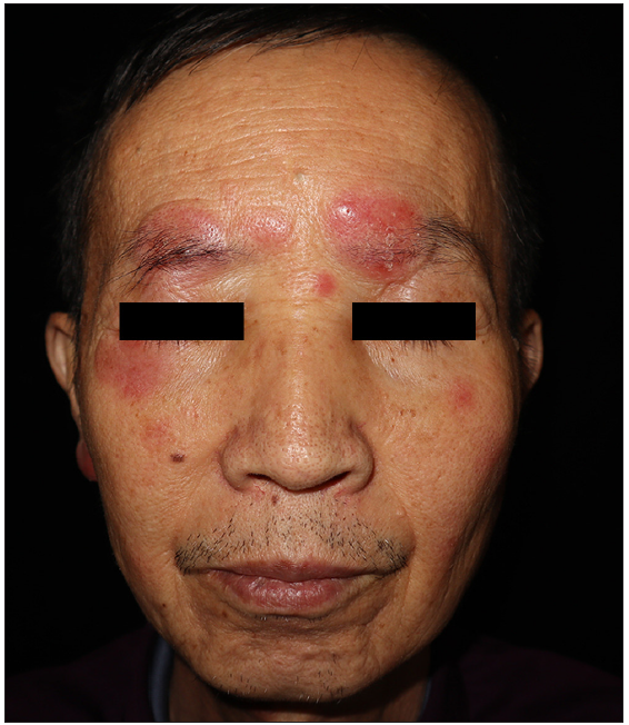
- Numerous, indurated erythematous nodules and plaques on the face.
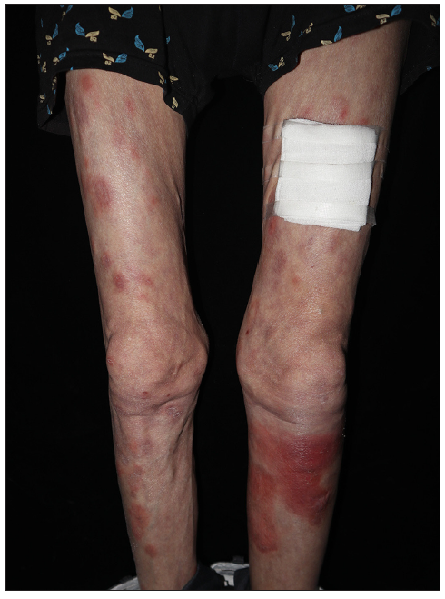
- Numerous, indurated erythematous nodules and plaques on the legs.
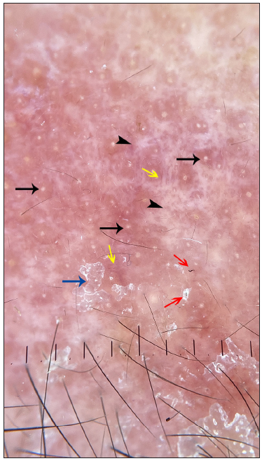
- Dermoscopy of the eyebrow lesion showed white areas (black arrowhead) surrounding the follicle plugging (black arrow) with a few short linear vessels (yellow arrow) at the periphery of follicles. Scales (blue arrow) and a few broken hairs (red arrow) are seen along with pinkish areas (polarising, 10x)
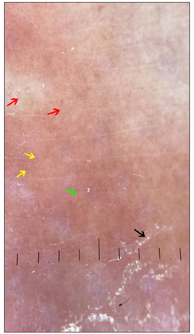
- Dermoscopy of the leg lesion revealed scales (black arrow), with distortion of pigment network (red arrow), and dotted (yellow arrow) or fine linear vessels (green arrow) in an orange-yellowish background on the right lower limb (polarising 10x).
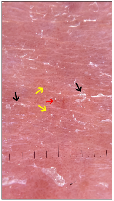
- Dermoscopy of the leg lesion observed a homogeneous dull‐red background, dotted (yellow arrow) and hairpin (red arrow) vessels with white scales (black arrow) on the left lower limb (polarising, 10x)
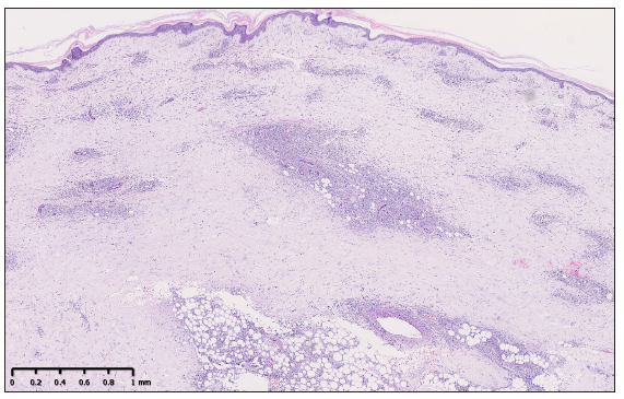
- Histopathological examination showed a dermal and subcutaneous infiltration of atypical lymphoid cells, with angiocentricity. (Haematoxylin and eosin, 40x)
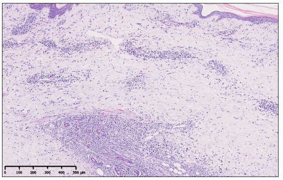
- Histopathological examination showed a dermal and subcutaneous infiltration of atypical lymphoid cells, with angiocentricity. (Haematoxylin and eosin, 100x)
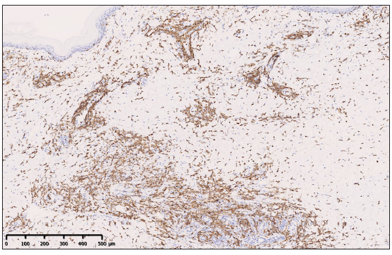
- Immunohistochemistry showed positive staining for CD3 of atypical cells (100x).
- Immunohistochemistry showed positive staining for CD8 of atypical cells (100x).
- Immunohistochemistry showed positive staining for CD56 of atypical cells (100x).
- Immunohistochemistry showed positive staining for granzyme B of atypical cells (100x).

- Epstein-Barr Virus stain (EBV) was demonstrated by in situ hybridisation (100x).
- The proliferative index (ki-67) was exceedingly high (80%) (100x).
Numerous studies have demonstrated that dermoscopy may be helpful in the assessment of cutaneous lymphoproliferative disorders, providing a correlation between clinical and histopathological examination. In our case, white areas and pinkish, orange-yellowish, or dull-red backgrounds correlate with dermal fibrosis and dermal atypical lymphocytic infiltrates, respectively. Polymorphous (dotted, fine linear, and hairpin) vessels are attributed to angiogenesis because of neoplastic processes. Broken hairs may indicate a potential injury to the hair shaft in the context of dense periadnexal atypical lymphocytic infiltrates.
The most commonly observed dermoscopic pattern of classical mycosis fungoides shows dotted, fine-short linear vessels resembling spermatozoa‐like structures with orange‐yellowish areas.3,4 The lower limb nodules of our patient had similar features. Moreover, the eyebrow lesion demonstrated white areas surrounding the follicle plugging with a few short linear vessels at the periphery of the follicles. In addition, scales and a few broken hairs are seen along with pinkish areas. These structures are most frequently associated with folliculotropic mycosis fungoides.5 On the other hand, primary cutaneous marginal zone B-cell lymphoma and primary cutaneous follicle centre lymphoma most commonly present with a salmon-coloured background and fine short/linear irregular/serpentine vessels.6 However, the dermoscopic features likely related to vessel dilatation and atypical lymphoid infiltrate around or within the affected vessel, as well as in the epidermis and the dermis, are not diagnostic. A histological work-up and a careful clinicopathologic correlation in the diagnostic approach to cutaneous lymphomas are needed.
In the present study, we would like to emphasise that the dermoscopic features of the primary cutaneous ENKTL of our patient are not specific and cannot be distinguished from classical or folliculotropic mycosis fungoides.
Declaration of patient consent
The authors certify that they have obtained all appropriate patient consent.
Financial support and sponsorship
Nil.
Conflicts of interest
There are no conflicts of interest.
Use of artificial intelligence (AI)-assisted technology for manuscript preparation
The authors confirm that there was no use of artificial intelligence (AI)-assisted technology for assisting in the writing or editing of the manuscript and no images were manipulated using AI.
References
- Cutaneous extranodal natural killer/T-cell lymphoma: A comparative clinicohistopathologic and survival outcome analysis of 45 cases according to the primary tumor site. J Am Acad Dermatol. 2014;70:1002-09.
- [CrossRef] [PubMed] [Google Scholar]
- Dermoscopy in the diagnosis of cutaneous lymphoma (Review) Exp Ther Med. 2022;23:377.
- [CrossRef] [PubMed] [PubMed Central] [Google Scholar]
- Dermoscopic patterns of early-stage mycosis fungoides in a Chinese population. Clin Exp Dermatol. 2019;44:169-75.
- [CrossRef] [PubMed] [Google Scholar]
- Morphologic features of cutaneous -cell lymphomas using dermoscopy and high frequency ultrasound. J Clin Med. 2020;10:17.
- [CrossRef] [PubMed] [PubMed Central] [Google Scholar]
- Early dermoscopic sign of folliculotropism in patients with mycosis fungoides. Dermatol Pract Concept. 2018;8:328-29.
- [CrossRef] [PubMed] [PubMed Central] [Google Scholar]
- Dermoscopy and the diagnosis of primary cutaneous B-cell lymphoma. J Eur Acad Dermatol Venereol. 2018;32:53-6.
- [CrossRef] [PubMed] [PubMed Central] [Google Scholar]


