Generic selectors
Exact matches only
Search in title
Search in content
Post Type Selectors
Search in posts
Search in pages
Filter by Categories
15th National Conference of the IAOMFP, Chennai, 2006
Abstract
Abstracts from current literature
Acne in India: Guidelines for management - IAA Consensus Document
Addendum
Announcement
Art & Psychiatry
Article
Articles
Association Activities
Association Notes
Award Article
Book Review
Brief Report
Case Analysis
Case Letter
Case Letters
Case Notes
Case Report
Case Reports
Clinical and Laboratory Investigations
Clinical Article
Clinical Studies
Clinical Study
Commentary
Conference Oration
Conference Summary
Continuing Medical Education
Correspondence
Corrigendum
Cosmetic Dermatology
Cosmetology
Current Best Evidence
Current Issue
Current View
Derma Quest
Dermato Surgery
Dermatopathology
Dermatosurgery Specials
Dispensing Pearl
Do you know?
Drug Dialogues
e-IJDVL
Editor Speaks
Editorial
Editorial Remarks
Editorial Report
Editorial Report - 2007
Editorial report for 2004-2005
Errata
Erratum
Focus
Fourth All India Conference Programme
From Our Book Shelf
From the Desk of Chief Editor
General
Get Set for Net
Get set for the net
Guest Article
Guest Editorial
History
How I Manage?
IADVL Announcement
IADVL Announcements
IJDVL Awards
IJDVL AWARDS 2015
IJDVL Awards 2018
IJDVL Awards 2019
IJDVL Awards 2020
IJDVL International Awards 2018
Images in Clinical Practice
In Memorium
Inaugural Address
Index
Knowledge From World Contemporaries
Leprosy Section
Letter in Response to Previous Publication
Letter to Editor
Letter to the Editor
Letter to the Editor - Case Letter
Letter to the Editor - Letter in Response to Published Article
LETTER TO THE EDITOR - LETTERS IN RESPONSE TO PUBLISHED ARTICLES
Letter to the Editor - Observation Letter
Letter to the Editor - Study Letter
Letter to the Editor - Therapy Letter
Letter to the Editor: Articles in Response to Previously Published Articles
Letters in Response to Previous Publication
Letters to the Editor
Letters to the Editor - Letter in Response to Previously Published Articles
Letters to the Editor: Case Letters
Letters to the Editor: Letters in Response to Previously Published Articles
Medicolegal Window
Messages
Miscellaneous Letter
Musings
Net Case
Net case report
Net Image
Net Images
Net Letter
Net Quiz
Net Study
New Preparations
News
News & Views
Obituary
Observation Letter
Observation Letters
Oration
Original Article
ORIGINAL CONTRIBUTION
Original Contributions
Pattern of Skin Diseases
Pearls
Pediatric Dermatology
Pediatric Rounds
Perspective
Presedential Address
Presidential Address
Presidents Remarks
Quiz
Recommendations
Regret
Report
Report of chief editor
Report of Hon : Treasurer IADVL
Report of Hon. General Secretary IADVL
Research Methdology
Research Methodology
Resident page
Resident's Page
Resident’s Page
Residents' Corner
Residents' Corner
Residents' Page
Retraction
Review
Review Article
Review Articles
Reviewers 2022
Revision Corner
Self Assessment Programme
SEMINAR
Seminar: Chronic Arsenicosis in India
Seminar: HIV Infection
Short Communication
Short Communications
Short Report
Snippets
Special Article
Specialty Interface
Studies
Study Letter
Study Letters
Supplement-Photoprotection
Supplement-Psoriasis
Symposium - Contact Dermatitis
Symposium - Lasers
Symposium - Pediatric Dermatoses
Symposium - Psoriasis
Symposium - Vesicobullous Disorders
SYMPOSIUM - VITILIGO
Symposium Aesthetic Surgery
Symposium Dermatopathology
Symposium-Hair Disorders
Symposium-Nails Part I
Symposium-Nails-Part II
Systematic Review and Meta-Analysis
Systematic Reviews and Meta-analyses
Systematic Reviews and Meta-analysis
Tables
Technology
Therapeutic Guideline-IADVL
Therapeutic Guidelines
Therapeutic Guidelines - IADVL
Therapeutics
Therapy
Therapy Letter
Therapy Letters
View Point
Viewpoint
What’s new in Dermatology
View/Download PDF

Translate this page into:

Observation Letter
87 (
2
); 278-280
doi:
10.25259/IJDVL_575_20

Dermoscopic findings of de novo syringocystadenoma papilliferum

Department of Dermatology All India Institute of Medical Sciences, Bhubaneswar, Odisha, India
Department of Pathology with Laboratory Medicine, All India Institute of Medical Sciences, Bhubaneswar, Odisha, India
Corresponding author: Dr. Biswanath Behera, Department of Dermatology, All India Institute of Medical Sciences, Bhubaneswar - 751 019, Odisha, India. biswanathbehera61@gmail.com
Licence
This is an open-access article distributed under the terms of the Creative Commons Attribution-Non Commercial-Share Alike 4.0 License, which allows others to remix, tweak, and build upon the work non-commercially, as long as the author is credited and the new creations are licensed under the identical terms.

How to cite this article: Dash S, Nayak AK, Sethy M, Palit A, Behera B. Dermoscopic findings of a de novo syringocystadenoma papilliferum. Indian J Dermatol Venereol Leprol 2021;87:278-80.

Sir,

Syringocystadenoma papilliferum is an uncommon, benign, hamartomatous adnexal tumor arising from pluripotent cells. The histology can exhibit either apocrine or eccrine differentiation. Although commonly associated with other benign adnexal tumours like sebaceous nevus, it can also occur de novo.1

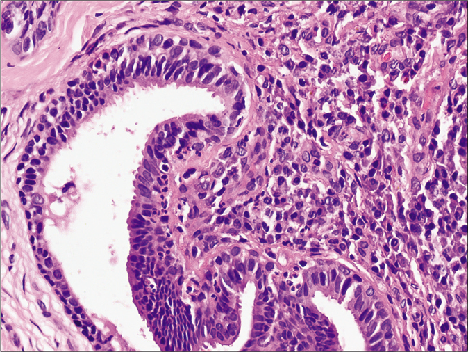
A 7-year-old healthy boy presented with multiple asymptomatic skin-colored, solid elevated lesions on his back since last 4 years. His personal and family history were unremarkable. Cutaneous examination demonstrated multiple, discrete, skin-colored to translucent, firm papules showing mid-back distribution. Most papules bore a central umbilication, while some coalesced to form plaques [Figure 1]. Remaining cutaneous, general and systemic examinations were within normal limits. Dermoscopy under polarized light (Dermlite, DL4, 10× magnification) revealed a pattern comprising of a central crater surrounded by white structureless area and/ or shiny white lines and an outer rim of brown structureless area [Figure 2a]. Occassional white rosettes (white dots, arranged in a square) and dotted vessels [Figure 2b] were identified. We considered syringocystadenoma papilliferum and molluscum contagiosum as differentials. Histopathology revealed hyperkeratosis, papillomatosis and dermal cystic invaginations [Figure 3a] lined by double-layered epithelium- an inner row of columnar cells and outer row of small cuboidal cells [Figure 3b]. We observed multiple luminal papillary projections, and the stroma was infiltrated with lymphocytes, histiocytes, plasma cells and sparse neutrophils. Few columnar cells showed decapitation secretion. The upper dermis showed dense cellular infiltration (lymphocytes > plasma cells), + while lower dermis demonstrated dilated apocrine glands. We made a diagnosis of syringocystadenoma pappiliferum and, referred the patient to the department of plastic surgery for complete surgical excision.

Figure 1:
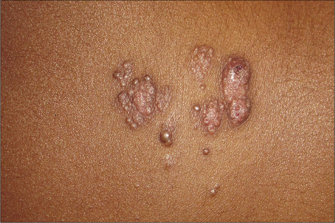
Multiple, discrete, skin-colored to translucent, firm papules on the mid-back with most having central umbilication and at places coalescing to form plaques.
Figure 2a:
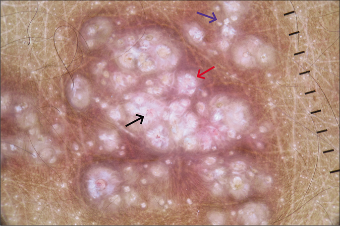
Dermoscopy (Dermlite, DL4, ×10) under polarized mode showing central crater surrounded by shiny white lines and an outer rim of brown structureless area (red arrow), white rosette (blue arrow) and dotted vessels (black arrow).
Figure 2b:
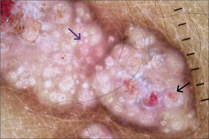
Dermoscopy (Dermlite, DL4, ×10) under polarized mode showing white rosette (blue arrow) and dotted vessels (black arrow).
Figure 3a:
Histopathology showing hyperkeratosis, papillomatosis, dermal cystic invaginations along with dense inflammatory cells in the upper dermis and dilated apocrine ducts in the lower dermis (H and E, ×100).
Figure 3b:
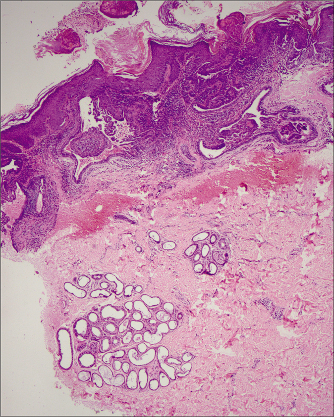
Histopathology showing cystic invaginations lined by an inner row of columnar cells and an outer row of small cuboidal cells. A few columnar cells showing decapitation secretion (H and E, ×400).

Syringocystadenoma papilliferum usually affects infants and children, while adolescents and adults constitute 15–30% cases. The head and neck region is affected most commomnly, in about 75% cases. Although benign, a rare transformation to basal cell, metastatic and ductal carcinoma has been reported. Three different clinical morphologies exist plaque, linear and solitary nodular type. The histogenesis of syringocystadenoma papilliferum is controversial and a hamartomatous origin from mature apocrine, eccrine or undifferentiated pluripotent cells is postulated.2

We found only eight previous reports describing the dermoscopic features of syringocystadenoma papilliferum, with only four having de novo origin. On dermoscopy, we observed, a central crater surrounded by white structureless area and/or shiny white lines in most papules, both discrete and coalescing. The central crater corresponds to the characteristic cystic invagination and the white structureless area represents the surrounding hyperplastic epidermis. A similar observation has been reported by Chauhan et al. and Zaballos et al., who described central depression and ulceration in syringocystadenoma papilliferum and correlated it histologically to cystic spaces opening to the skin.1,3 We were unable to find any previous report depicting shiny white structures and lines, including rosettes, features of polarized mode. Haspeslagh et al. described smaller (0.1–0.2 mm) and larger (0.3–0.5 mm) rosettes, and attributed them to cross-polarization of concentric horn material in follicular and eccrine ducts at the infundibular level of biopsy and concentric perifollicular fibrosis, respectively.4 In our case, the rosettes may be attributed to perifollcicular or perieccrine ductal or pericrater (polarizing) keratinisation.1 Zaballos et al. have reported a polymorphic vascular pattern and correlated the vascular structures to the fibrovascular core of papillary projections; which corroborates our finding of dotted vessels inside the craters.3 Unlike in our case, Lombardi and colleagues reported a yellowish hue representing the underlying nevus sebaceus.5

Molluscum contagiosum is the closest differential of our case, dermoscopically characterized by the co-existence of orifices, polylobular yellowish-white material or uniform homogenous color in the center, vascular structures (crown, punctiform, radial and mixed flower) and white rosettes.6 This vascular pattern is restricted to the amorphous structure, and never crosses the core in contast to syringocystadenoma papilliferum, in which vascular structures may involve the central crater, as in our case. Another close differential diagnosis is linear unilateral basaloid follicular hamartoma, dermoscopically distinguished by brown-gray globules and dots, in focus dots, brown linear and arciform structures, crown vessels, short fine telangiectasias, spoke wheel-like structures without a central dark point, white structureless areas with telangiectasias and keratotic plug.7

To conclude, the dermoscopic pattern consisting of a central crater surrounded by a white homogenous area, representing cystic invagination and surrounding epidermal hyperplasia respectively, can be a valuable diagnostic clue for de novo syringocystadenoma papilliferum. Additionally, we have described two novel dermoscopic features- shiny white lines and rosette.

Declaration of patient consent

The authors certify that they have obtained all appropriate patient consent.

Financial support and sponsorship

Nil.

Conflicts of interest

There are no conflicts of interest.

References

  1. , , , . Dermoscopy of a rare case of linear syringocystadenoma papilliferum with review of the literature. Dermatol Pract Concept. 2018;8:33-8.
    [CrossRef] [PubMed] [Google Scholar]
  2. , , , . Syringocystadenoma papilliferum: A case series and review of the literature in Indian context. Hell J Surg. 2014;86:98-101.
    [CrossRef] [Google Scholar]
  3. , , , , , , et al. Dermoscopy of tumours arising in naevus sebaceous: A morphological study of 58 cases. J Eur Acad Dermatol Venereol. 2015;29:2231-7.
    [CrossRef] [PubMed] [Google Scholar]
  4. , , , , , , et al. Rosettes and other white shiny structures in polarized dermoscopy: Histological correlate and optical explanation. J Eur Acad Dermatol Venereol. 2016;30:311-3.
    [CrossRef] [PubMed] [Google Scholar]
  5. , , , , , , et al. Dermoscopy of syringocystadenoma papilliferum. Australas J Dermatol. 2018;59:e59-61.
    [CrossRef] [PubMed] [Google Scholar]
  6. , . Rosette: An additional in vivo dermoscopic finding in molluscum contagiosum. Indian J Pediatr Dermatol. 2019;20:349-50.
    [CrossRef] [Google Scholar]
  7. , , . Clinicopathological and dermoscopic features in a case of linear and unilateral basaloid follicular hamartoma. Indian Dermatol Online J. 2019;10:710-3.
    [CrossRef] [PubMed] [Google Scholar]

Fulltext Views
2,950

PDF downloads
2,166
View/Download PDF
Download Citations
BibTeX
RIS
Show Sections