Translate this page into:
Dermoscopic findings of Spitz nevus on acral volar skin
2 Department of Pathology, Seoul National University College of Medicine, Seoul National University, Seoul, South Korea
Correspondence Address:
Je-Ho Mun
Department of Dermatology, Seoul National University College of Medicine, 101 Daehak-Ro, Jongno-Gu, Seoul 110-744
South Korea
| How to cite this article: Montenegro Jaramillo SE, Jo G, Darmawan CC, Lee C, Mun JH. Dermoscopic findings of Spitz nevus on acral volar skin. Indian J Dermatol Venereol Leprol 2019;85:629-632 |
Sir,
Spitz nevus is a benign melanocytic neoplasm usually affecting children and young adults. It can appear as a reddish-pink or brownish-black lesion, predominantly distributed on the face, neck, or lower extremities while acral location is rare. Dermoscopy is a useful tool in the differential diagnosis of acral melanocytic tumors. However, data regarding their dermoscopic features on the acral volar skin is scarce. We report two cases of acral Spitz nevus on the glabrous skin, with dermoscopic and histopathologic findings.
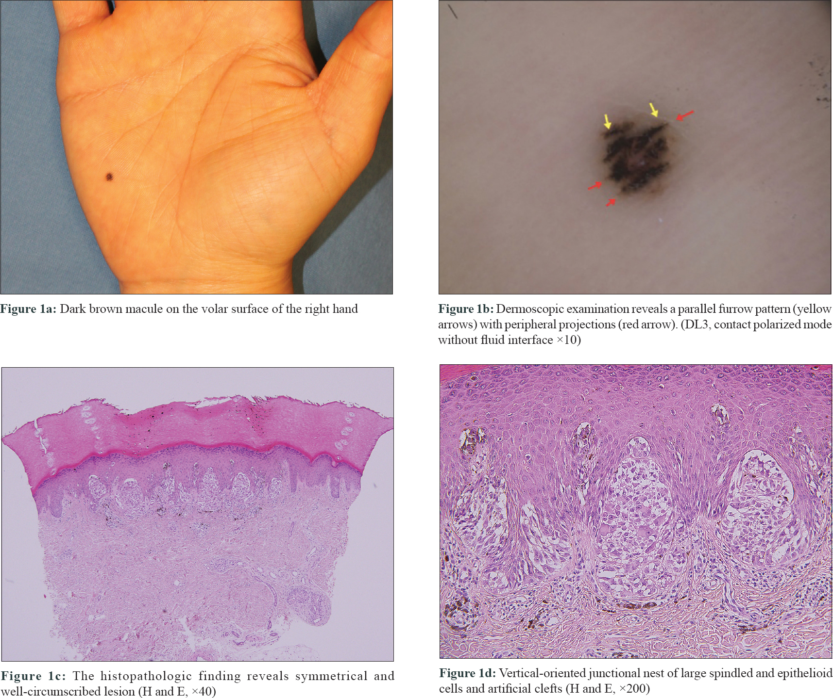
A 23-year-old woman was referred to our department with a pigmented lesion on her right palm since last 5 years. Physical examination revealed a dark brown macule sized about 3 × 2 mm. [Figure - 1]a. Dermoscopy using contact polarized mode without fluid interface (DL3 equipment; 3Gen, San Juan Capistrano, CA, USA) revealed a furrow pattern (yellow arrows) with peripheral projections (red arrow) [Figure - 1]b. Histopathologic examination demonstrated a symmetric and well-circumscribed lesion composed of melanocytic nests. Several vertically-oriented junctional nests composed of large, heavily melanised spindled and epithelioid melanocytes with abundant cytoplasm were observed on higher magnification. Artificial clefts were conspicuous between the nests and epidermal cells. [Figure - 1]c and [Figure - 1]d. Immunohistochemical tests showed positive staining for Melan A (MART-1) and a low Ki-67 proliferation index. Based on the clinical and histopathologic findings, a diagnosis of acral Spitz nevus was made.
 |
| Figure 1: |
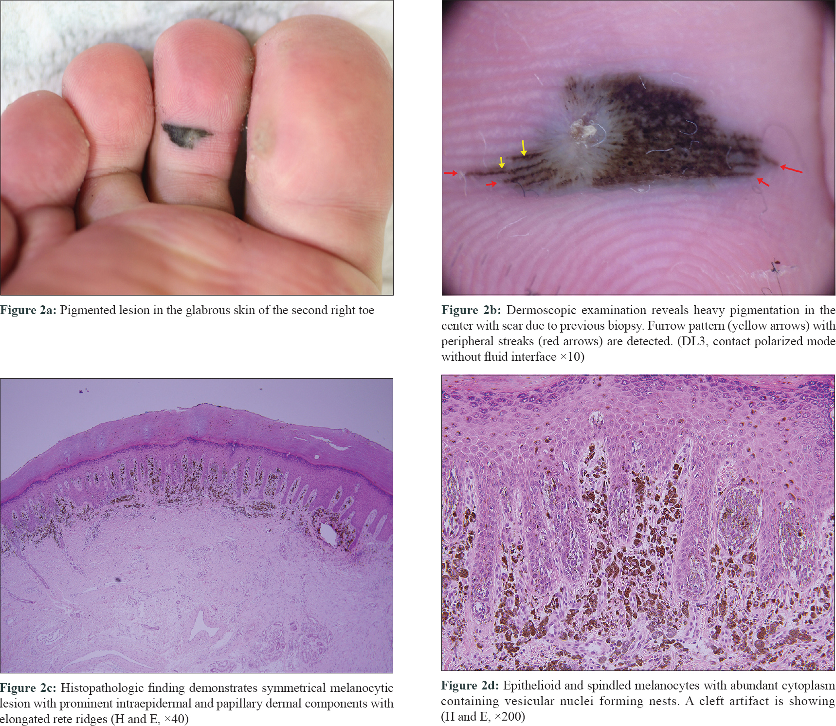
A 23-year-old man was referred to us with a 13 × 14 mm sized pigmented patch on the plantar surface on the second toe for the last 18 months [Figure - 2]a, showing atypical melanocytic features on skin biopsy. Dermoscopic evaluation with polarized mode showed heavy central pigmentation with a scarred area due to a previous biopsy. In the margins, a parallel furrow pattern (yellow arrows) with peripheral streaks (red arrows) was detected [Figure - 2]b. We excised the lesion with 3-mm free margins and repaired it with a bilobed flap. Histopathologic analysis revealed junctional melanocytic proliferation along with elongated dermal rete ridges. Under high magnification, heavily pigmented epithelioid and spindled melanocytes containing vesicular nuclei were observed to form nests. Clefting was detected around them; however, mitotic figures were absent [Figure - 2]c and [Figure - 2]d. We diagnosed acral spitz nevus, based on these findings.
 |
| Figure 2: |
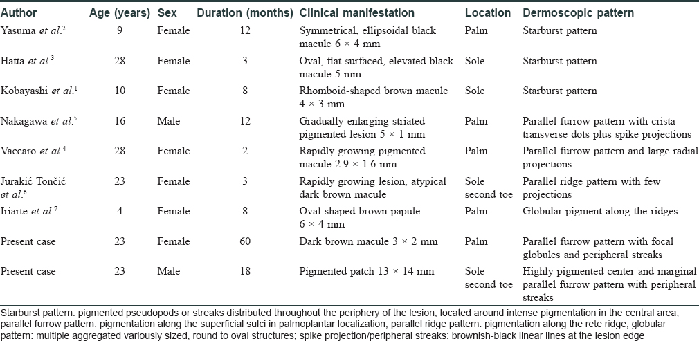
Palmo-plantar spitz nevus is rare. Spitz nevus on glabrous skin has been reported to comprise about 2% of all Spitz nevi.[1] We have found only seven dermoscopic reports of acral spitz nevi on in the literature [Table - 1].[1],[2],[3],[4],[5],[6],[7] Starburst pattern [1],[2],[3], parallel furrow pattern with peripheral dots and projections [4],[5] and a parallel ridge pattern with few peripheral globules [6] have been reported in three, two and a single case respectively. Recently, globular pigment along the ridges has been reported.[7] In our first case, we detected a furrow pattern with peripheral streaks while our second case demonstrated a heavily pigmented central area with marginal parallel furrows and streaks. Parallel ridge pattern is a diagnostic dermoscopic feature of acral melanoma. However, most of the reported cases including the current ones (78%) presented benign patterns. Therefore, dermoscopy can be a useful ancillary technique for acral Spitz nevus. However, more cases are needed to validate their dermoscopic features.

Spitz nevus is a unique melanocytic tumor, often resembling malignant melanoma clinically, thus requiring a high index of suspicion. Clinical history (young age), histopathologic and dermoscopic evaluation are necessary for accurate diagnosis.[4] In our cases, the dermoscopic findings differed from the typical parallel ridge pattern of acral melanoma. Histologic features like asymmetric melanocytic distribution, atypical hyperchromatic melanocytes with Pagetoid spread, and high mitotic figures with increased ki-67 index are suggestive of acral melanoma. In contrast, the symmetric proliferation of medium-sized to large epitheloid or spindle-shaped melanocytes with retraction artefact (clefting), Kamino bodies, and dermal maturation without cellular or nuclear pleomorphism suggest acral spitz nevus.[8] In addition, clinical information is essential to differentiate acral lentiginous melanoma from Spitz nevi as the former usually occurs in elderly patients. In our case, the characteristics of both lesions indicated acral Spitz nevi.
In conclusion, we report two rare cases of acral Spitz nevi with both histopathologic and dermoscopic findings. Further reports of dermoscopic patterns of acral spitz nevus will help in definining the characteristics of this entity.
Declaration of patient consent
The authors certify that they have obtained all appropriate patient consent forms. In the form the patients have given their consent for their images and other clinical information to be reported in the journal. The patients understand that their names and initials will not be published and due efforts will be made to conceal their identity, but anonymity cannot be guaranteed.
Financial support and sponsorship
Nil.
Conflicts of interest
There are no conflicts of interest.
| 1. |
Kobayashi H, Oishi K, Miyake M, Nishijima C, Kawashima A, Kobayashi H, et al. Spitz nevus on the sole of the foot presenting with transepidermal elimination. Dermatol Pract Concept 2014;4:41-3.
[Google Scholar]
|
| 2. |
Yasuma A, Hara H, Hukuda N, Terui T. Usefulness of dermoscopy for diagnosing pigmented spitz nevus occurring on the glabrous skin. J Eur Acad Dermatol Venereol 2006;20:1362-3.
[Google Scholar]
|
| 3. |
Hatta N, Arai M, Makino S. Dermoscopic findings of pigmented Spitz nevus of the sole. J Dermatol 2012;39:1048-9.
[Google Scholar]
|
| 4. |
Vaccaro M, Borgia F, Cannavò SP. Dermoscopy of pigmented variant of acral spitz nevus. J Am Acad Dermatol 2015;72:S11-2.
[Google Scholar]
|
| 5. |
Nakagawa K, Kishida M, Okabayashi A, Shimizu N, Taguchi M, Kinoshita R, et al. Spitz nevus on the palm with crista transverse dots/dotted lines revealed by dermoscopic examination. J Dermatol 2015;42:649-50.
[Google Scholar]
|
| 6. |
Jurakić Tončić R, Bradamante M, Ferrara G, Štulhofer-Buzina D, Petković M, Argenziano G. Parallel ridge dermoscopic pattern in plantar atypical Spitz nevus. J Eur Acad Dermatol Venereol 2018;32:e101-e102.
[Google Scholar]
|
| 7. |
Iriarte C, Rao B, Haroon A, Kirkorian AY. Acral pigmented spitz nevus in a child with transepidermal migration of melanocytes: Dermoscopic and reflectance confocal microscopic features. Pediatr Dermatol 2018;35:e99-102.
[Google Scholar]
|
| 8. |
Wiedemeyer K, Guadagno A, Davey J, Brenn T. Acral spitz nevi: A clinicopathologic study of 50 cases with immunohistochemical analysis of P16 and P21 expression. Am J Surg Pathol 2018;42:821-7.
[Google Scholar]
|
Fulltext Views
4,305
PDF downloads
1,363


