Translate this page into:
Dermoscopy of arteriovenous hemangioma
Corresponding author: Dr. Chi-Ling Lin, Department of Dermatology, Kaohsiung Medical University Hospital, 100 Tzyou 1st Road, Kaohsiung 807, Taiwan. 930307@kmuh.org.tw
-
Received: ,
Accepted: ,
How to cite this article: Hu SC, Lin CL. Dermoscopy of arteriovenous hemangioma. Indian J Dermatol Venereol Leprol 2021;87:298-9.
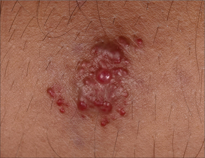
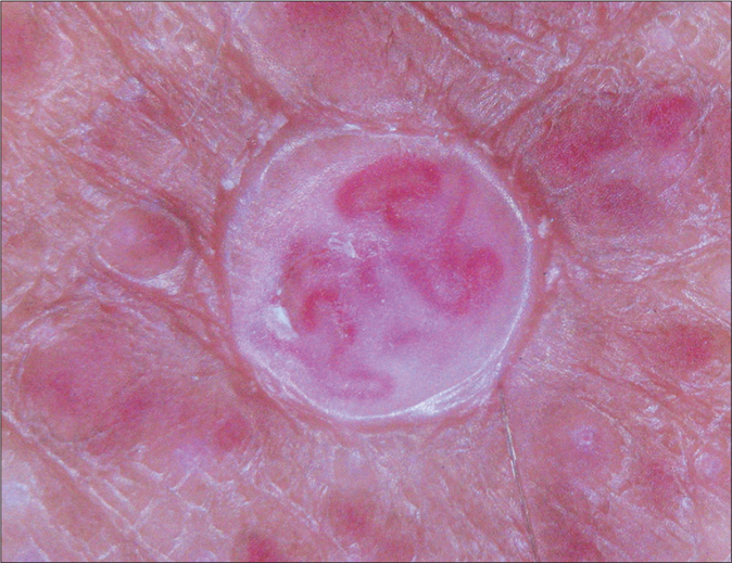
A 30-year-old man presented with multiple erythematous papules coalescing into a 1.5-cm plaque on his left forearm [Figure 1a], which had been present for more than 10 years. On dermoscopic examination (DermLite Foto (3Gen, Dana Point, California), magnification 20×, no contact fluid used), most of the papules were characterized by homogeneous erythematous background, while some of the larger papules showed telangiectatic serpentine vessels (long, winding vascular structures) [Figure 1b]. A skin biopsy showed numerous thick-walled arteries and veins in the dermis. A diagnosis of arteriovenous hemangioma (malformation) was made. Previously, the most common dermoscopic feature of arteriovenous hemangioma was found to be non-arborizing telangiectasia on a reddish background.1 Therefore, dermoscopy may be a valuable tool for the preoperative diagnosis of this vascular malformation.

- The patient presented with grouped, erythematous papules on his left forearm

- Dermoscopic examination revealed homogeneous erythematous background and telangiectatic serpentine vessels
Financial support and sponsorship
Nil.
Conflicts of interest
There are no conflicts of interest.
References
- Dermoscopy of arteriovenous tumour: A morphological study of 39 cases. Australas J Dermatol. 2018;59:e253-7.
- [CrossRef] [PubMed] [Google Scholar]


