Translate this page into:
Detection of pemphigus autoantibodies in healthy relatives of Turkish patients with pemphigus
2 Department of Pathology, Istanbul University Cerrahpasa Medical Faculty, Istanbul, Turkey
Correspondence Address:
Ozgur Emek Kocaturk
Bozova Sk. Bayirbasi Apt. No: 4/10 Mecidiyekoy, Sisli, Istanbul
Turkey
| How to cite this article: Kavala M, Kocaturk OE, Demirkesen C, Can B, Zindanci I, Turkoglu Z. Detection of pemphigus autoantibodies in healthy relatives of Turkish patients with pemphigus. Indian J Dermatol Venereol Leprol 2007;73:240-242 |
Abstract
Background: Pemphigus autoantibodies have been reported in healthy relatives of pemphigus patients suggesting a genetic predisposition in the pathogenesis of the disease. Aims: To test for the presence of pemphigus autoantibodies in healthy relatives of Turkish patients of pemphigus. Methods: The study group comprised 45 pemphigus patients, 75 unaffected family members and 47 healthy individuals in the control group. Direct and indirect immunofluorescence techniques were performed to determine the presence of pemphigus autoantibodies. Results: By indirect immunofluorescence staining, circulating pemphigus autoantibodies were found in 26.7% of the relatives and in only two of the controls ( P value = 0.0001). A direct immunofluorescence technique revealed positive results in three (4%) of the relatives and none of the controls. Conclusion: The presence of pemphigus autoantibodies in clinically healthy relatives indicates that genetic predisposition is necessary but not sufficient for the development of clinical disease.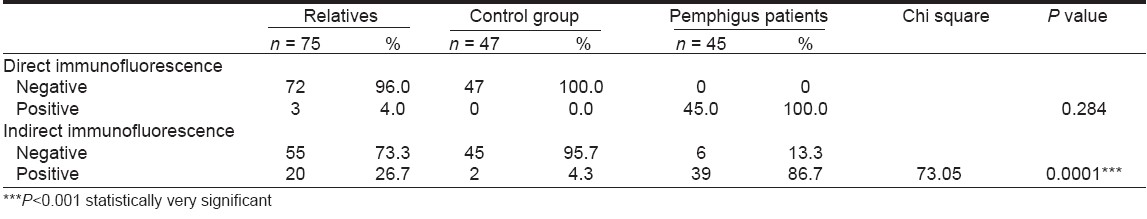
Pemphigus is a life-threatening autoimmune disease, which is characterized by intraepidermal bulla and acantholysis and is mediated by IgG autoantibodies. [1] The autoantibodies are thought to be a result of exogeneous and endogenous factors. [2]
Evidences like the frequent occurrence of pemphigus in some ethnic groups such as Ashkenazi Jews and Japanese, [3] its association with the presence of some HLA alleles, [4],[5] occurrence of familial pemphigus cases [2],[6] and reports of circulating pemphigus autoantibodies in the healthy relatives of pemphigus patients [1],[2],[7],[8] indicate a role for genetic predisposition in its pathogenesis. We evaluated the frequency of pemphigus autoantibodies in healthy relatives of patients with pemphigus in a Turkish population.
Methods
Our study included 45 pemphigus patients who were referred to our outpatient clinic between October 1998 and January 2004. The diagnosis of pemphigus was made on the basis of clinical, histological and immunofluorescence examinations. The severity of the clinical disease was assessed to be mild, moderate and severe. For direct immunofluorescence (DIF) examination, a 4 mm punch biopsy specimen was taken from the left arm of the relatives and of the controls. The biopsy specimens were frozen in liquid nitrogen and stored in at -70°C until use. The specimens were cut by cryostat into 5 µ thick sections. After being rinsed with phosphate-buffered saline solution (PBS) for 15 minutes, the slides were incubated with anti-human IgG and complement (Dako-polyclonal rabbit) at a dilution of 1/100 in PBS, pH 7.2 at room temperature for 45 minutes. The slides were rinsed three times for ten minutes in PBS.
For indirect immunofluorescence (IIF) tests reactivity of fluorescence intensity was assessed between the titers of 1/10-1/160 for intercellular IgG deposits. For IIF, the blood was drawn from both groups at the time of biopsy. The sera were evaluated by a standard IIF technique using human skin as the substrate.
The analyses were performed using the statistical programme SPSS for Windows 10.0 ((Statistical Package for Social Sciences). Chi-square, Fischer′s exact test, Mann Whitney U and ANOVA tests were used for the comparison of results. P < 0.05 was considered as statistically significant. Prior approval of the local ethics committee was taken and informed consent was obtained from participants.
Results
Thirty-two of the patients were women (71.1%) and 13 were men (28.9%). Their mean age was 52.3 years (range = 24-74 years). The pemphigus subtypes were as follows: four patients with pemphigus erythematosus (8.9%), one patient with pemphigus foliaceus (2.2%), one patient with pemphigus herpetiformis (2.2%) and 39 patients with pemphigus vulgaris (86.7%). The disease activity was mild in four (8.9%), moderate in 35 (77.8%) and severe in six (13.3%) patients.
The study also included 75 first-degree healthy relatives of which 32 were women (42.7%) and 43 were men (57.3%) with a mean age of 29.6 years (range = 7-77 years). A control group consisting of 47 healthy individuals, of which 28 were women (59.6%) and 19 were men (40.4%), the mean age being 36.4 years (range = 12-74 years), was also included.
DIF revealed negative results in 72 (96%) of the relatives while it was positive in three (4%). IIF was also positive for two of these three relatives. There were no positive DIF results in the control group. IIF was positive in 20 (26.7%) relatives. Of the 47 individuals in the control group, 45 (95.7%) showed negative IIF results while two (4.3%) showed positive results. In six (13.3%) of the pemphigus patients, IIF gave negative results [Table - 1].
There were no statistically significant differences in the frequency of positive DIF results between the relatives of the pemphigus patients and the control group. The frequency of IIF positivity in the relatives′ group was found to be significantly high when compared to that of the control group (26.7% vs 4.3%) ( P < 0.001). No statistically significant correlation was found between clinical severity of the disease in the patients′ group and the frequency of IIF positivity in the relatives′ group ( P >0.05).
Discussion
Pemphigus vulgaris (PV) is strongly associated with some major histocompatibility (MHC) alleles. The alleles that are frequently found are DR4 and DQ8 in Jewish patients and DR6 and DQ5 in nonJewish patients. [9] The relationship between pemphigus and MHC has been well demonstrated but only a few cases of familial pemphigus have been reported. Beutner and Chorzelski [10] found only one case of familial PV in their series of 234 cases. Reohr et al. [11] studied the MHC of two siblings with PV by restriction fragment polymorphism methods and found that both of them had DR4 and DQW3.2 alleles. Though their father had the same haplotypes, he did not develop clinical signs of PV.
In many studies, low titers of pemphigus autoantibodies were found in the first-degree relatives of pemphigus patients. Ahmed et al found low levels of PV autoantibodies in 48% of 120 healthy first-degree relatives of 31 PV patients and they linked the presence of these autoantibodies to HLA-DR4 or DR6 haplotypes of the patients. [9] Kricheli et al. detected pemphigus autoantibodies in 15% of 55 healthy relatives of 25 pemphigus patients by using the IIF technique. However, none of the 56 controls had PV-IgG in their sera. [8] The antibody titers of the relatives ranged from 1/10 to 1/160. Brandsen et al searched for the presence of pemphigus autoantibodies by DIF, IIF and immunoblotting techniques in 21 first-degree relatives of 12 pemphigus patients and a control group consisting of 30 individuals. [2] They found a higher incidence of PV-IgG (71%) by using the three techniques. These IgGs were demonstrated to bind the epidermal cells in only five (25%) of the relatives. There was no positivity found in the control group by any of the methods used.
In a series of 12 Italian pemphigus patients, a significant number of the 67 first-degree relatives had PV-IgG as determined by IIF (31%) and immunoblotting methods (47%). [7] The antibody titers were between 1/20 and 1/80. In another survey of 56 first-degree relatives of 37 pemphigus cases, the incidences of pemphigus autoantibodies were 42.8 and 19.8% by IIF and ELISA methods, respectively. [1] In the present study, we found pemphigus autoantibodies in 26.7% of the relatives. These results were between 15-31.1%, a range that has been detected in previous studies. [7],[8] The range of antibody titers (1/10 to 1/160) detected in our study, was consistent with the range reported in Kricheli′s study. [8] On the other hand, the rate of positivity detected by DIF (4%) in our study was significantly lower than the rate that Brandsen et al determined (25%). This situation may be due to the small sample size that they had used. [2]
Although there is a genetic predisposition in pemphigus, the question as to why the relatives of the patients do not show clinical signs of the disease remains unanswered. Kricheli et al suggest that the antibody titers detected in the sera of the relatives were not high enough to bind the pemphigus antigens in the skin and induce acantholysis. Thus, the disease may be triggered by infections or drugs. [8] These authors also found that the distribution of PV IgG1-3 subclasses were similar among patients and their relatives but PV IgG4 was higher in patients and lower in relatives. This finding may indicate the role of PV IgG subclasses in the pathogenesis of the disease.
In almost all of these studies, circulating antibodies have been well detected by the IIF technique while the DIF results were usually negative. This suggests an immunologic or mechanical barrier that interferes with the interaction between circulating IgG and the skin. This barrier may be demolished by drugs, malignancies, viral infections or burns, which may result in clinical disease. The variability of the susceptibility of keratinocytes or the penetrability of the epidermis may thus determine who develops clinical disease and who remains asymptomatic with circulating pemphigus antibodies. [2] On the basis of the previous and present studies, we conclude that though immunogenetic factors constitute a basic part in the pathogenesis of pemphigus, they are not sufficient for the development of the disease. Details of interactions between genetic and environmental factors need further illumination.
| 1. |
Torzecka JD, Narbutt J, Sysa-Jedrzejowska A, Waszczykowska E, Lukamowicz J, Pas HH. Detection of pemphigus autoantibodies by IIF and ELISA tests in patients with pemphigus vulgaris and foliaceus and in healthy relatives. Med Sci Monit 2003;9:CR528-33.
[Google Scholar]
|
| 2. |
Brandsen R, Frusic-Zlotkin M, Lyubimov H, Yunes F, Michel B, Tamir A, et al. Circulating pemphigus IgG in families of patients with pemphigus: Comparison of indirect immunofluorescence, direct immunofluorescence and immunoblotting. J Am Acad Dermatol 1997;36:44-52.
[Google Scholar]
|
| 3. |
Martel P, Joly P. Pemphigus: Autoimmune diseases of Keratinocyte's adhesion molecules. Clin Dermatol 2001;19:662-74.
[Google Scholar]
|
| 4. |
Krain LS, Terasaki PI, Newcomer VD, Mickey MR. Increased frequency of HLA-A10 in pemphigus vulgaris. Arch Dermatol 1973;108:803-5.
[Google Scholar]
|
| 5. |
Szafer F, Brautbar C, Tzfoni E, Frankel G, Sherman L, Cohen I, et al. Detection of disease spesific restriction fragment length polymorphism in pemphigus vulgaris linked to the DQW1 and DQW3 alleles of the HLA-D region. Proc Natl Acad Sci USA 1987;84:6542-5.
[Google Scholar]
|
| 6. |
Brenner S, Dorfman B, Himelfarb M. Familial pemphigus vulgaris. Dermatologica 1985;171:38-40.
[Google Scholar]
|
| 7. |
Mohimen A, Narula M, Ruocco V, Pisani M, Ahmed AR. Presence of the autoantibody in healthy relatives of Italian patients with pemphigus vulgaris. Arch Dermatol Res 1993;285:176-7.
[Google Scholar]
|
| 8. |
Krichelli D, David M, Frusic-Zlotkin M, Goldsmith D, Rabinov M, Sulkes J, et al. The distribution of pemphigus vulgaris-IgG subclasses and their reactivity with desmoglein 3 and 1 in pemphigus patients and their first degree relatives. Br J Dermatol 2000;143:337-42.
[Google Scholar]
|
| 9. |
Ahmed AR, Mohimen A, Yunis EJ, Mirza NM, Kumar V, Beutner EH, et al. Linkage of pemphigus vulgaris to the major histocompatibility complex in healthy relatives of patients. J Exp Med 1993;177:419-24.
[Google Scholar]
|
| 10. |
Beutner EH, Chorzelski TP. Studies on etiologic factors in pemphigus. J Cutan Pathol 1976;3:67-74.
[Google Scholar]
|
| 11. |
Reohr PB, Mangklabruks A, Janiga AM, DeGroot LJ, Banjasuratwong Y, Soltani K. Pemphigus vulgaris in siblings: HLA-DR4 and HLA-DQW3 and susceptibility to pemphigus. J Am Acad Dermatol 1992;27:189-93.
[Google Scholar]
|
Fulltext Views
940
PDF downloads
437


![[Table - 1]](#tbl_ijdvl_2007_73_4_240_32889_1.jpg)