Translate this page into:
Disfiguring morphology of tuberous sclerosis
Correspondence Address:
Sujay Khandpur
Department of Dermatology and Venereology, All India Institute of Medical Sciences, New Delhi
India
| How to cite this article: Khandpur S, Gupta S. Disfiguring morphology of tuberous sclerosis. Indian J Dermatol Venereol Leprol 2011;77:403-404 |
Sir,
Tuberous sclerosis complex (TSC) is characterized by the development of hamartomas in various organs. Approximately one in 8000 adults and one in 6000 newborns is affected by TSC. [1] Skin lesions occur in 60-70% of the cases and are a major cause of morbidity. We present an interesting case of TSC with florid cutaneous manifestations that produced significant disfigurement and physical morbidity.
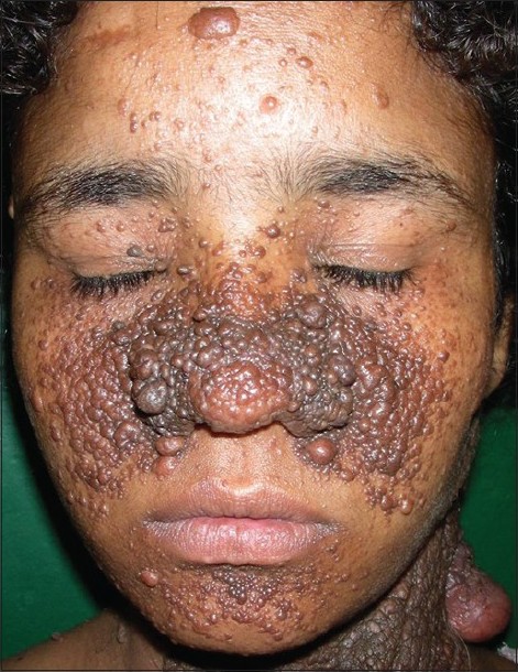
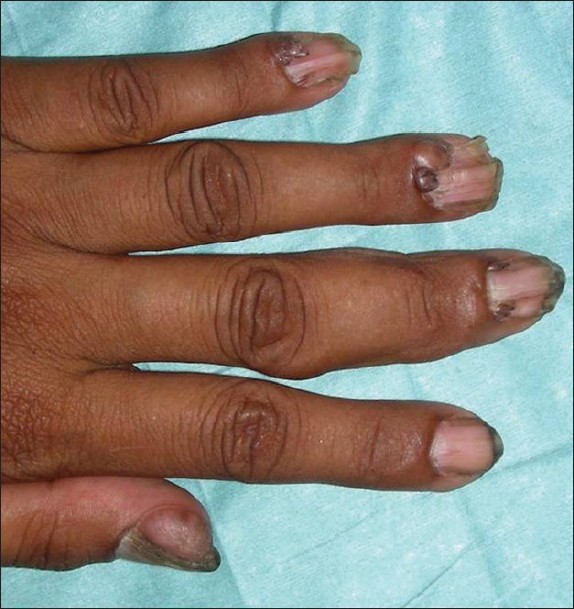
A 25-year-old girl presented with malodorous plaques over the face and neck since 2 years of age, papules in the oral cavity since 5-6 years and brownish papules and plaques over the nail folds of almost all fingers and toes since 8-10 years. There was history of unprovoked violent behavior since 1-2 years with delayed mental milestones, but no history of seizures, abdominal pain, urinary complaints, blurring of vision or backache. The patient was brought to the skin outpatient department (OPD) as she was cosmetically distressed by the skin lesions. She was a product of non-consanguineous marriage. There was no family history of tuberous sclerosis. The patient′s general physical and systemic examinations were normal. Cutaneous examination revealed multiple, dark brown to erythematous, discrete to coalescent papules and plaques, some pedunculated, suggestive of angiofibromas, predominantly over the central face, retroauricular area, left forearm and bilateral thighs [Figure - 1]. There was a diffuse brown, firm bosselated plaque studded with nodules, predominantly over the left side of the neck and upper chest. Multiple forehead plaques and many brownish plaques suggestive of shagreen patch were present over the lower back. Peri- and subungual fibromas (Koenen′s tumors), few being globose or having verrucous surface, involving all finger and toe nails and producing longitudinal grooves in the nail plates, were present [Figure - 2]. The oral cavity showed multiple (12-15), skin-colored to hyperpigmented, 1-3-mm-sized sessile and pedunculated papules over the lower labial mucosa and tip of the tongue, suggestive of mucosal fibromas.
 |
| Figure 1: Numerous erythematous to hyperpigmented large and small angiofibromas over the face and neck |
 |
| Figure 2: Periungual fibromas involving all fingers |
Investigations were normal except urine analysis, which revealed hematuria and pyuria. X-ray of the hands showed sclerosis in all proximal and middle phalanges of the fingers bilaterally. Ultrasound and computed tomography (CT) scan of the abdomen showed multiple angiomyolipomas in both kidneys. Chest X-ray showed minimal bilateral pleural effusion with few nodules and cysts in the bilateral lower lobes. CT scan head showed calcified subependymal nodules in the bilateral lateral ventricles and right cerebellar hemispheres and tubers in the left posterior parietal cortex. ECG and echocardiography were normal. The patient was referred to a plastic surgeon for excision of the unsightly facial tumors.
TSC is one of the common genodermatosis encountered in the dermatology OPD. However, this case is interesting in view of the grotesque mucocutaneous manifestations.
Tuberous sclerosis is an autosomal-dominant disorder with variable expression and a high rate of spontaneous mutations. Consequently, the clinical picture is inconstant, with large differences in manifestations and course of the disease, even in members of the same family. The existence of a gene of major effect that produces disease, coupled with modifier genes that influence expressivity, may explain this variability. [2]
Among adults with TSC, skin lesions take on a greater significance and, for about 15%, are a regular problem. Facial angiofibromas and periungual fibromas may be a source of recurrent bleeding and chronic infection, and may become painful. For others, they represent a significant cosmetic concern.
Angiofibromas usually appear at the age of 6 years, and are most common around puberty. The severity of the angiofibromas varies from small, barely noticeable papules, to large, disfiguring bulbous nodules. They have been reported to produce generalized overgrowth of tissues of the forearm and wrist, causing pain and reduced function. [3] A very disfiguring case of TCS manifesting as large ulcerated angiomatous tumor covering the entire right side of the face with numerous angiofibromas over the head and neck, associated fibromatous scalp lesions and shagreen patches, which required excision and grafting for facial lesion, has been reported. [4]
In a cross-sectional study of cutaneous features in TSC, of the 131 affected individuals, 96% exhibited skin signs, 81% patients had facial angiofibromas at age up to 14 years while only 54% had shagreen patches till this age. [3] Hypomelanotic macules were identified in 61% of the patients, of which 89% had more than one and 40% had more than five. Ungual fibromas were multiple in 75% of the patients, and were more common on the toenails (90%) than on the fingernails. Extensive skin tags were present on the neck, axilla, upper back or inguinal area in 6% of the patients. Oral manifestations included gingival fibromas in 36% of the patients and involved the anterior gingivae and, occasionally, buccal mucosa or under false teeth. Multiple or large glossal hamartomas requiring surgical excision have been reported.
Many of the cutaneous manifestations of TSC have also been found in multiple endocrine neoplasia (MEN) type 1 syndrome. Of 32 MEN1 patients, 88% had multiple (range 2-50) facial angiofibromas and 72% had discrete papular collagenomas. [5] Angiofibromas in MEN1 patients were smaller and fewer than in severely affected patients of TSC, and were often observed on the upper lip and vermilion border, areas spared in TSC. Also observed were cafι au lait macules in 38%, confetti-like hypopigmented macules in 6% and multiple gingival papules in 6%.
| 1. |
Napolioni V, Curatolo P. Genetics and molecular biology of tuberous sclerosis complex. Curr Genomics 2008;9:475-87.
[Google Scholar]
|
| 2. |
Boesel CP, Paulson GW, Kosnik EJ, Earle KM. Brain hamartomas and tumors associated with tuberous sclerosis. Neurosurgery 1979;4:410-7.
[Google Scholar]
|
| 3. |
Webb DW, Clarke A, Fryer A, Osborne JP. The cutaneous features of tuberous sclerosis: A population study. Br J Dermatol 1996;135:1-5.
[Google Scholar]
|
| 4. |
Alvarez E. Unusual cutaneous manifestations of tuberous sclerosis: Case report. Plast Reconstr Surg 1967;40:153-6.
[Google Scholar]
|
| 5. |
Darling TN, Skarulis MC, Steinberg SM, Marx SJ, Spiegel AM, Turner M. Multiple facial angiofibromas and collagenomas in patients with multiple endocrine neoplasia type 1. Arch Dermatol 1997;133:853-7.
[Google Scholar]
|
Fulltext Views
8,669
PDF downloads
2,945


