Translate this page into:
Eruptive hypomelanosis in a child, a new viral exanthem
2 Department of Dermatology, Division of Medicine, King Edward Memorial Hospital, Pune, Maharashtra, India
3 JC School of Public Health, The Chinese University of Hong Kong and The Prince of Wales Hospital, Hong Kong, China
Correspondence Address:
Vijay Zawar
Skin Disease Center, Shreeram Sankul, Opp. Hotel Panchavati, Vakilwadi, Nashik - 422 001, Maharashtra
India
| How to cite this article: Zawar V, Bharatia P, Chuh A. Eruptive hypomelanosis in a child, a new viral exanthem. Indian J Dermatol Venereol Leprol 2016;82:85-86 |
Sir,
Eruptive hypomelanosis is a newly described paraviral exanthem reported from India.[1],[2]
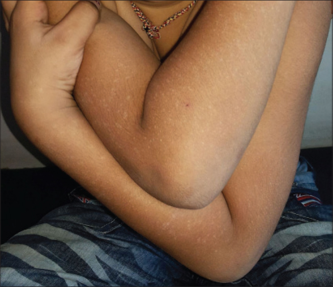
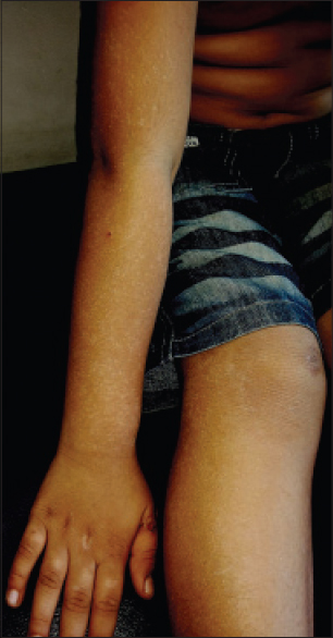
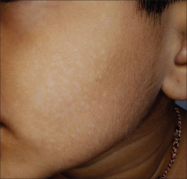
A 6-year-old boy presented with sudden onset of progressive, asymptomatic crops of well-demarcated, symmetrical, hypomelanotic macules ranging in size from 0.1 to 0.5 cm [Figure - 1],[Figure - 2],[Figure - 3]. The eruption appeared in crops and began on the forearms and then spread to legs and thighs and face over a period of 1 week. Most lesions were non-scaly and discrete. Some closely situated macules showed confluence. After the progressive eruption involved all four extremities and the cheeks, the rash appeared to stop progressing further. The trunk, palms, soles and mucosa were spared. He had a transient episode of coryza and mild fever for 10 days before onset of the eruption for which no treatment was sought. Apart from mild congestion of the throat, the remainder of the physical examination was normal.
 |
| Figure 1: Multiple, symmetric, sharply demarcated and dense crops of eruptive hypopigmented macules of acute onset and of nearly similar shape and size over the extensor forearms and arms |
 |
| Figure 2: Similar lesions over the extensor right forearm, arm, thigh and leg. Similar eruptions were also present on contralateral side |
 |
| Figure 3: Similar lesions on the left cheek with confl uence of some lesions. Similar eruptions were also present on the right cheek |
His baseline laboratory investigations including complete blood counts, urinalysis and blood sugar levels were normal. There was no evidence of pityriasis versicolor as revealed by Wood's lamp examination and potassium hydroxide preparation done on scrapings of the lesions. His family members declined a skin biopsy. We prescribed topical white petroleum alone and closely monitored him. The hypopigmentation gradually faded 1 month after onset of the eruption.
We considered the differential diagnoses of pityriasis versicolor, pityriasis alba, progressive macular hypomelanosis and post-inflammatory hypopigmentation. However, none of these diagnoses was supported by the investigations and clinical observations. The onset as hypopigmented macules without any preceding inflammation negates the likelihood of post-inflammatory hypopigmentation. Progressive macular hypopigmentation was also unlikely because of the short course of disease.[3]
Eruptive hypomelanosis is likely to be caused by a viral infection because of (i) a prodromal coryzal phase for 1 to 2 weeks preceding the onset of the eruption, (ii) the eruptive nature with successive crops of lesions, (iii) the fairly uniform sizes of lesions, (iv) spontaneous resolution without active intervention and (v) relatively young age of the children affected, thus indicating a primary infection rather than endogenous reactivation of a virus.[1],[2] We believe that it is a paraviral exanthem, meaning that a single viral etiology is yet to be identified.
Lipsker and Saurat classified paraviral exanthems into three categories, (i) clinically well-defined eruptions having many causes with at least one clearly primary infection or endogenous reactivation of viruses such as Gianotti-Crosti syndrome and papulo-purpuric gloves and socks syndrome, (ii) typical and recognizable eruptions with viral infections or reactivations suspected as a triggering cause, but which do not have a yet identified cause such as asymmetric periflexural exanthem and eruptive pseudoangiomatosis and (iii) clinically very distinctive eruptions having long been classified as paraviral in dermatological nosology, but not due to solidly documented direct viral-related cytopathogenic effects such as pityriasis rosea.[4] In this classification, eruptive hypomelanosis could be placed in the second category because the viral cause is suspected but unconfirmed. However, as no virus is currently known to be associated with eruptive hypomelanosis, we do not have an evidence-based long-term follow-up strategy.
It is not clear why are there no reports of this condition from other ethnic groups and other countries apart from India, from where there are two reports by our group describing ten patients.[1],[2] One reason might be that a hypopigmented rash would be more prominently noted in darker-skinned races. Another reason could be the self-limited course of the rash. Thus, many children with eruptive hypomelanosis might not be brought to medical attention. It is likely that a significant proportion of such children are seen only by primary care physicians and not referred to dermatologists or pediatricians because the rash is asymptomatic and self-resolving. Finally, specialists may not be aware of this new clinical entity because of a paucity of published reports.
We look forward to more clinicians reporting this rash. We recommend epidemiological studies of this novel paraviral exanthem to establish the infectious nature of the eruption. While we were unable to carry them out because of resource constraints, investigations that may be helpful include (i) lesional biopsy for histopathology, immunohistochemical staining and electron microscopy, (ii) specimens from multiple body sites (lesion, plasma and peripheral blood mononuclear cells) for polymerase chain reaction to detect DNA various viruses and real-time polymerase chain reaction to determine the virus load, and (iii) acute and convalescent sera for serological investigations to be conducted in parallel to detect immunoglobulin M (relatively non-specific), seroconversion (the gold standard for primary viral infections) and significant rise of titers of antibodies against viruses. Data from these investigations would help us delineate this novel exanthem and may provide insights into appropriate management.
Financial support and sponsorship
Nil.
Conflicts of interest
There are no conflicts of interest.
| 1. |
Zawar V, Bharatia P, Chuh A. Eruptive hypomelanosis: A novel exanthem associated with viral symptoms in children. JAMA Dermatol 2014;150:1197-201.
[Google Scholar]
|
| 2. |
Chuh A, Bharatia P, Zawar V. Eruptive hypomelanosis occurring in a young child – A brief report. Pediatr Dermatol 2015;32. [In press].
[Google Scholar]
|
| 3. |
Relyveld GN, Menke HE, Westerhof W. Progressive macular hypomelanosis: An overview. Am J Clin Dermatol 2007;8:13-9.
[Google Scholar]
|
| 4. |
Lipsker D, Saurat JH. A new concept: Paraviral eruptions. Dermatology 2005;211:309-11.
[Google Scholar]
|
Fulltext Views
9,576
PDF downloads
2,855


