Translate this page into:
Eruptive lichen planus in a child with celiac disease
Correspondence Address:
Amrinder J Kanwar
Department of Dermatology, Venereology and Leprology, Postgraduate Institute of Medical Education and Research, Chandigarh - 160012
India
| How to cite this article: De D, Kanwar AJ. Eruptive lichen planus in a child with celiac disease. Indian J Dermatol Venereol Leprol 2008;74:164-165 |
 |
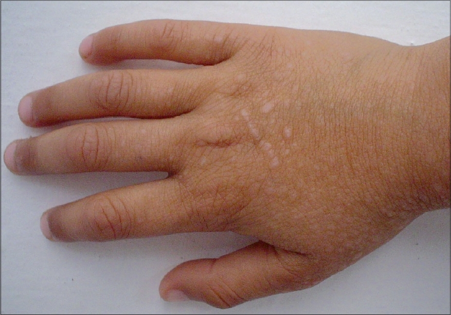
| Figure 1: Violaceous papules with koebnerization over dorsum of hand |
 |
| Figure 1: Violaceous papules with koebnerization over dorsum of hand |
Sir,
A 6-year-old boy had diarrhea and abdominal distension since the age of 1 year. He was investigated and was found to have celiac disease. His complete blood count, liver and renal function tests and fasting blood sugar were within normal limits. His IgG anti-gliadin antibody and IgA anti-transglutaminase antibodies were negative, and duodenal biopsy showed evidence of celiac disease (Marsh grade 2). He was started on gluten-free diet and his celiac disease improved. Two months before seeking dermatology consultation, he started developing showers of itchy violaceous papules all over his body. He was not on any active medication for celiac disease except the gluten-free diet. On examination, he had multiple violaceous monomorphic papules all over with evidence of koebnerization [Figure - 1]. A punch biopsy from one of the papules showed classical changes of lichen planus (LP). He was given 20 mg prednisolone per day orally with which the skin lesions healed within 3 weeks.
Celiac disease is a chronic T-cell-mediated enteropathy against ingested gluten in genetically predisposed individuals. [1] Unlike previously thought, recent epidemiological studies have shown celiac disease to be a common disorder affecting 1 in 250 persons in the US and UK populations. [2] It has been associated with HLA-DQ2, expressed in more than 80% of patients or HLA-DQ8. [3]
Many extra-intestinal associations have been observed in celiac disease patients, including dermatological manifestations. Most of the dermatological manifestations are secondary to nutritional deficiency. A few autoimmune dermatological manifestations have also been described, most important among which is dermatitis herpetiformis. Others include linear IgA bullous disease, vitiligo, alopecia areata, dermatomyositis, etc. However, there is only one report of a single case of erosive oral LP with celiac disease. [4]
It is surprising that association of cutaneous LP and celiac disease has never been reported previously, although both are not very uncommon and are T-cell-mediated autoimmune disorders. Perhaps, the different genetic linkages of LP (HLA-DQ1) and celiac disease (HLA-DQ2 and HLA-DQ8) may explain this. This report will increase awareness of this association amongst dermatologists and pediatricians so that more such instances may be identified in the future.
| 1. |
Schuppan D. Current concepts of celiac disease pathogenesis. Gastroenterology 2000;119:234-42.
[Google Scholar]
|
| 2. |
Fasano A, Catassi C. Current approaches to diagnosis and treatment of celiac disease: An evolving spectrum. Gastroenterology 2001;120:636-51.
[Google Scholar]
|
| 3. |
Green PH, Jabri B. Celiac disease. Lancet 2003;362:383-91.
[Google Scholar]
|
| 4. |
Fortune F, Buchanan JA. Oral lichen planus and coeliac disease. Lancet 1993;341:1154-5.
[Google Scholar]
|
Fulltext Views
6,189
PDF downloads
1,469


