Translate this page into:
Erythematosquamous plaque on a closed ostomy site
Corresponding author: Dr. Ruben Linares-Navarro, Department of Dermatology, University Hospital of León, Calle Altos de Nava S/N, León, Spain. rubenlinaresnavarro@hotmail.com
-
Received: ,
Accepted: ,
How to cite this article: Linares-Navarro R, Olmos-Nieva CC, García-Sanz M, Gutierrez-Carrillo G, Rodriguez-Prieto MA. Erythematosquamous plaque on a closed ostomy site. Indian J Dermatol Venereol Leprol. 2024;90:91-2. doi: 10.25259/IJDVL_47_2023
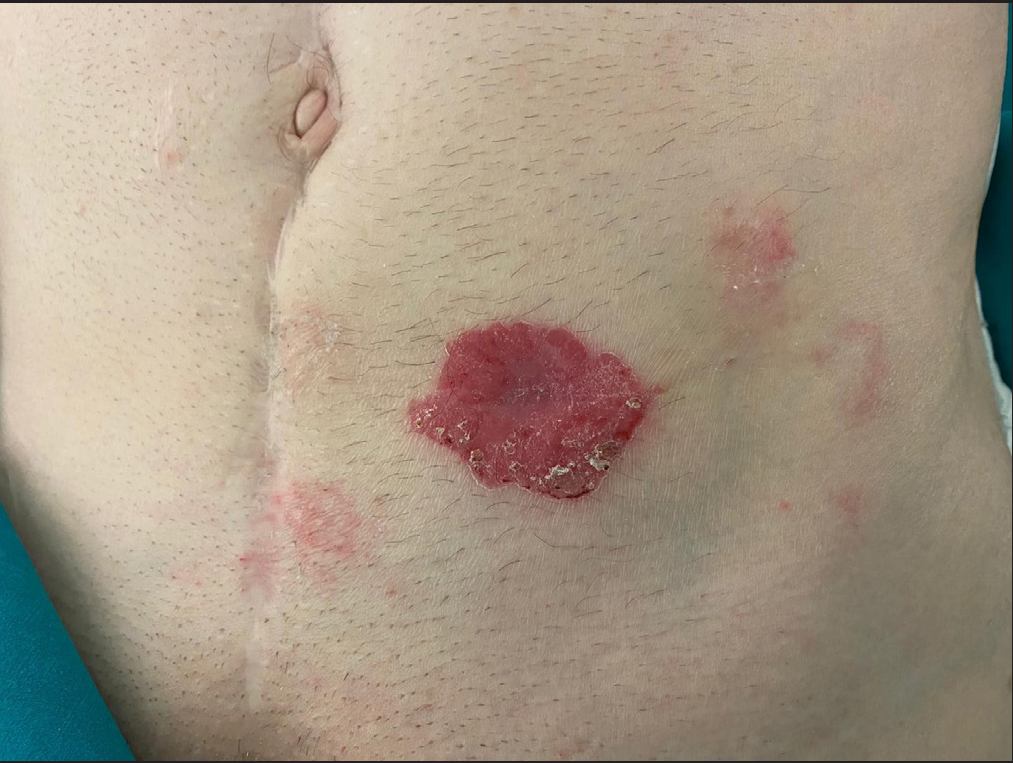
A 68-year-old man with a history of urothelial carcinoma of bladder with extension to the left ureter was treated with laparoscopic radical cystectomy, partial left ureterectomy and nephrostomy. Five years later, he presented with an erythematosquamous plaque on a previous ostomy site that had already been closed, located in the left abdominal region [Figure 1].
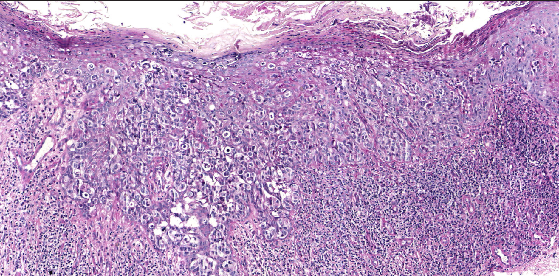
Biopsy of the lesion revealed permeation of the epidermis by tumour cells with vesicular nuclei, visible nucleoli and large vacuolated cytoplasm that were scattered in single cells and in small groups in the epidermis. In the superficial dermis there was an intense inflammatory infiltrate consisting mainly of lymphocytes [Figure 2].

- Erythematosquamous plaque in a previous ostomy site that had already been closed, located in the left abdominal region.

- Permeation of the epidermis by tumour cells with vesicular nuclei, visible nucleoli and large vacuolated cytoplasm. In the superficial dermis there was an intense inflammatory infiltrate (haematoxylin and eosin stain, x100).
Question
What is your diagnosis?
Diagnosis
Pagetoid extension of urothelial carcinoma from ureter to skin.
Discussion
Cases of secondary Paget’s disease in relation to urothelial carcinoma are well known. However, cases of urothelial carcinoma with pagetoid extension to the skin by direct contiguity are very rare. After a rigorous search of the literature, we were only able to find two cases, both in periostomal skin.1,2
Melicow et al. first described the pagetoid variant of urothelial carcinoma in situ in 1952.3 In our case it was considered as a pagetoid extension by contiguity of urothelial carcinoma, due to the location of the cutaneous lesion outside the typical areas of Paget’s disease, coinciding with the area of adhesion of the ureter to the abdominal wall.
However, the neoplasm in the residual ureter and the one present years ago in the bladder did not present a pagetoid pattern. This phenomenon has also been described in the case reported by Ito et al.,1 who proposed that pagetoid cells may have developed de novo from urothelial carcinoma in the ureter and subsequently spread to the skin.
Immunohistochemistry can help us discriminate between primary and secondary Paget’s disease, the latter corresponding to a pagetoid extension of an extracutaneous neoplasm. The positivity of CK 7, CK20 and P63 is characteristic of secondary Paget’s disease and makes it necessary to rule out the presence of malignant internal neoplasm.4,5
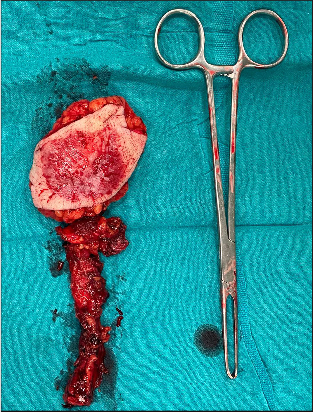
In view of the findings, it was decided to perform a laparoscopic distal left ureterectomy and resection of the skin lesion in the same surgical time [Figure 3].

- En bloc resection of the residual ureter and skin was performed.
Histological analysis of the ureter was compatible with a high-grade solid urothelial carcinoma infiltrating the skin with pagetoid extension. Immunohistochemistry was equivalent to that of the skin lesion: positive in neoplastic cells for Cytokeratin 7 (CK7), Cytokeratin 20 (CK20), P63 and GATA-3. Gross cystic disease fluid protein 15 (GCDFP-Q15) was negative.
The patient experienced clinical resolution. Adjuvant therapy was discouraged due to the patient’s history of renal insufficiency. One year after surgery, he is clinically and radiologically stable.
As clinicians, we should be aware of the possibility of recurrence in patients with a history of urinary tract cancer, in the presence of pagetoid lesions in the periostomal area.
Declaration of patient consent
Patient’s consent not required as patients identity is not disclosed or compromised.
Financial support and sponsorship
Nil.
Conflict of interest
There are no conflicts of interest.
References
- Peristomal pagetoid spread of urothelial carcinoma of the ureter. Rare Tumors. 2013;5:162-4.
- [CrossRef] [PubMed] [PubMed Central] [Google Scholar]
- A Case of secondary extramammary Paget’s disease around the cutaneous stoma after radical cystectomy. Hinyokika Kiyo. 2017;63:381-6.
- [CrossRef] [PubMed] [Google Scholar]
- Intra-urothelial cancer: carcinoma in situ, Bowen’s disease of the urinary system: discussion of thirty cases. J Urol. 1952;68:763-72.
- [CrossRef] [PubMed] [Google Scholar]
- The use of cytokeratins 7 and 20 in the diagnosis of primary and secondary extramammary Paget’s disease. Br J Dermatol. 2000;142:243-7.
- [CrossRef] [PubMed] [Google Scholar]
- Immunohistochemistry of p63 in primary and secondary vulvar Paget’s disease. Pathol Int. 2008;58:648-51.
- [CrossRef] [PubMed] [Google Scholar]


