
Translate this page into:
Erythematous plaque on left axillary area
-
Received: ,
Accepted: ,
How to cite this article: Liu XK, Li J. Erythematous plaque on left axillary area. Indian J Dermatol Venereol Leprol 2022;88:260-1.
80-year-old male presented with 6-month history of asymptomatic, erythematous lesion on the left axillary region. The patient had been treated for a fungal infection and dermatitis, but did not show any amelioration. Cutaneous examination revealed a 1.5 × 2 cm well-marginated infiltrated plaque on the left axilla without erosion [Figure 1]. Axillary lymphnodes were normally palpable. Ultrasound and mammography did not reveal evidence of breast cancer and no internal malignancy was detected by further studies, including computed tomography and serum tumor markers. The skin biopsy revealed epidermal infiltration by enlarged atypical cells with pale-staining cytoplasm and distinctive nuclei; findings that were in agreement with Paget’s disease. An immunohistochemical evaluation revealed tumor cells that were positive for carcinoembryonic antigen and cytokeratin 7 and negative for HMB-45 and Melan-A. We made the final diagnosis of primary extramammary Paget’s disease of the axilla. Wide local excision was performed on the patient and no recurrence was established at the 1-year follow-up visit
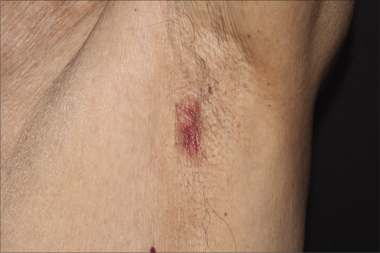
- A 1.5 cm × 2 cm well-marginated erythematous plaque on the left axilla
Declaration of patient consent
The authors certify that they have obtained all appropriate patient consent forms. In the form, the patient has given his consent for his images and other clinical information to be reported in the journal. The patient understand that name and initials will not be published and due efforts will be made to conceal identity, but anonymity cannot be guaranteed.
Financial support and sponsorship
Nil.
Conflicts of interest
There are no conflicts of interest.


