Translate this page into:
Erythrodermic scabies in a systemic sclerosis patient
Corresponding author: Dr. Anand Mannu, Department of Dermatology, Armed Forces Medical College, Pune, Maharashtra, India. anandstanley09@gmail.com
-
Received: ,
Accepted: ,
How to cite this article: Das P, Mannu A, Vasudevan B, Krishnan LP, Priya S. Erythrodermic scabies in systemic sclerosis patient. Indian J Dermatol Venereol Leprol. doi: 10.25259/IJDVL_384_2024
Dear Editor,
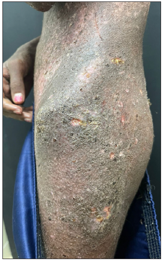
A 22-year-old female presented with salt and pepper pigmentation scattered across her body for the past one year associated with moderate itching. Over the last 6 months, she experienced bluish discolouration of her fingers upon cold exposure, diffuse tightening of the skin, which progressed from her digits to the entire body, restricted mouth opening, swallowing difficulties, and a history of breathlessness on exertion. Pallor, positive Barnett neck sign and Ingram sign could be elicited. Her body mass index (BMI) was 15. Investigations revealed elevated Anti Nuclear Antibody Anti nuclear antibodies (ANA) titres, positive Anti-SCL-70 and Anti-SS-A antibodies and a restrictive pattern on spirometry. Her electrolytes, ECG and 2D Echo were normal. She was diagnosed as a case of systemic sclerosis and was started on nifedipine, methotrexate, folic acid, bosentan, pantoprazole and domperidone, along with lifestyle modifications. During the hospital stay, she developed gradually progressive pruritis associated with scaling initially over the legs which progressed to involve all parts of the body (>80% body surface area) resulting in erythroderma [Figures 1a, 1b and 1c]. While evaluating for the causes of erythroderma, we found the attender developed recent onset itching in the finger web spaces which clinched the diagnosis of crusted scabies. A skin scraping from the patient revealed scabies mites when examined under a microscope [Figure 2, Supplementary video]. Both patient and attender showed improvement on treatment with topical permethrin and oral ivermectin.

- Extensive scaling over the trunk and flexures.

- Thick crusting, scaling and hyperpigmentation over left axilla.

- Crusting, scaling and excoriations over waist and thigh regions

- Scabies mite in liquid paraffin under microscope (10x)
Norwegian Scabies (NS) or Crusted Scabies (CS) is a rare and extremely infectious condition caused by Sarcoptes scabiei. The name “Norwegian” was derived from the description by Danielssen and Boeck of a type of scabies in which a huge number of mites were present in lepers.1 CS is characterised by an infestation of up to millions of mites and extensive hyperkeratotic plaques with yellow-green crusts, most commonly on the flexures, torso, extremities, face and scalp. Itching varies from asymptomatic to severe itching in a few patients. The mean incubation period is 3–4 weeks and crusted plaques appear after 8–12 weeks.2 Conventional scabies differ from CS by harbouring less than 15 mites, clinically by the presence of burrows and papules with intense itching at night. Immunologically, it differs by immunity switch from Th1 to Th2 with predominant CD8+ T cells, minimal CD4+ T cells and absence of B cells in the dermis. Also, IL 1β, TGF β, IL 4, IgE, IgG, IgG1, IgG3, IgG4 and IgA levels are elevated while IRAP, IL-10 and TNF-α levels are found to be reduced.3 CS are typically found in immunocompromised patients with HIV, human T-lymphotropic virus (HTLV), adult T-cell lymphoma, leprosy, epidermolysis bullosa, IgA deficiency, Langerhans cell histiocytosis, neutropenia, myelodysplasia, pregnancy, debilitated individuals with dementia, malnutrition and mental retardation.4 In our case, the patient’s immunocompromised state from medication and malnourishment raised her likelihood of developing CS.
CS is commonly misdiagnosed as psoriasis, adverse drug reactions, hyperkeratotic eczema or contact dermatitis and is generally diagnosed after a median of 3–7.8 months (range 16 months) after the initial symptoms. Diagnosis is by examination of scrapings under a microscope. Treatment includes oral ivermectin (200 ug/kg on days 1, 2, 8, 9 and 15) and topical 5% permethrin applied every 2–3 days for 1–2 weeks or till cure.5 By the end of the first week of treatment, our patient’s symptoms had improved. As in our case, there is a markedly higher chance of transmission to other individuals in contact since there is an enormous mite load and the patient should be isolated till cure. In hospital settings, the diagnosis of CS necessitates increased vigilance and once confirmed, stringent control measures to stop the spread of the disease need to be ensured.6
Acknowledgement
The authors would like to thank Dr Senkadhir Vendhan for taking the images and video.
Declaration of patient consent
The authors certify that they have obtained all appropriate patient consent.
Financial support and sponsorship
Nil.
Conflicts of interest
There are no conflicts of interest.
Use of artificial intelligence (AI)-assisted technology for manuscript preparation
The authors confirm that there was no use of artificial intelligence (AI)-assisted technology for assisting in the writing or editing of the manuscript and no images were manipulated using AI.
References
- Norwegian scabies: Rare cause of erythroderma. Indian Dermatol Online J. 2015;6:52-4.
- [CrossRef] [PubMed] [PubMed Central] [Google Scholar]
- Crusted (Norwegian) scabies: An under-recognized infestation characterized by an atypical presentation and delayed diagnosis. J Eur Acad Dermatol Venereol. 2016;30:483-5.
- [CrossRef] [PubMed] [Google Scholar]
- New insights into disease pathogenesis in crusted (Norwegian) scabies: The skin immune response in crusted scabies. Br J Dermatol. 2008;158:1247-55.
- [CrossRef] [PubMed] [Google Scholar]
- Norwegian scabies management after prolonged disease course: A case report. Int J Surg Case Rep. 2019;61:180-3.
- [CrossRef] [PubMed] [PubMed Central] [Google Scholar]
- Erythrodermic scabies in an immunocompetent patient. JAAD Case Rep. 2022;29:112-5.
- [CrossRef] [PubMed] [PubMed Central] [Google Scholar]
- An outbreak of scabies in a long-term care facility: The role of misdiagnosis and the costs associated with control. Infect Control Hosp Epidemiol. 2006;27:517-8.
- [CrossRef] [PubMed] [Google Scholar]


