Translate this page into:
Evaluation of the antioxidant status in vitiligo patients in Kashmir valley-A hospital based study
2 Department of Biochemistry, Government Medical College Srinagar, University of Kashmir, J and K, India
Correspondence Address:
Iffat Hassan
Postgraduate Department of Dermatology, STD and Leprosy, Government Medical College Srinagar, University of Kashmir, J and K
India
| How to cite this article: Hassan I, Hussain S, Keen A, Hassan T, Majeed S. Evaluation of the antioxidant status in vitiligo patients in Kashmir valley-A hospital based study. Indian J Dermatol Venereol Leprol 2013;79:100-101 |
Sir,
Vitiligo is an idiopathic, acquired, circumscribed hypomelanotic skin disorder, characterized by milky white patches of different sizes and shapes. It affects about 1-2% of the world population. [1] Several theories have been proposed about the pathogenesis of vitiligo, but the precise cause behind melanocyte destruction remains unknown. Theories regarding the destruction of melanocytes include autoimmune mechanisms, cytotoxic mechanisms, an intrinsic defect of melanocytes, and neural mechanisms. One of the hypotheses that have been recently proposed is the antioxidant deficiency theory. [2]
The present study was designed to evaluate the various antioxidant parameters of vitiligo patients in by measuring their plasma Superoxide dismutase (SOD) activity as well as plasma Glutathion reductase (GSH), Malondialdehyde (MDA) and Total antioxidative potential (AOP) levels.
This study was a hospital based study conducted in the Department Of Dermatology, STD and Leprosy of SMHS Hospital (associated teaching hospital of Government Medical College Srinagar). It included thirty eight patients with vitiligo (20 males and 18 females). Patients who had received any immunosuppressive drugs were excluded from the study. Other exclusion criteria were those patients who had an associated autoimmune disease or a malignancy, and who were smokers. An equal number of age and sex matched controls (20 males and 18 females) were selected as controls.
Fasting venous blood samples (5ml) were obtained from patients with vitiligo and healthy controls and were centrifuged to obtain plasma samples, which were then stored at a temperature of -20°C until analyzed.
GSH level was estimated by the method of Moron MS et al, [3] SOD activity was estimated by the method described by Kono Y et al. [4] Total amount of lipid peroxidation products was estimated using the thiobarbituric acid (TBA) method, which measures the TBA reactive products chiefly the MDA. [5] Total AOP was assessed by modified method of Ozturk et al. [6]
Statistical analysis of the data was performed by using Statistical Package for Social Sciences (SPSS Version 17).
The present study included 38 cases of vitiligo patients, and an equal number of age and sex matched controls. In the study group of 38 patients, there were 20 (52.63%) males and 18 (47.37%) females, with a male to female ratio of 1: 1.11. The age of our patients ranged from 5-50 years. The control group comprised of 38 (20 males and 18 females) healthy age matched subjects.
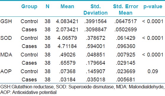
The plasma GSH levels were lower in cases as compared to controls (p-value < 0.0001). The plasma levels of SOD and MDA were found to be higher in cases as compared to controls (p-value < 0.0001). There was no statistically significant difference in the plasma AOP levels between the cases and controls (p-value was 0.09) [Table - 1].

The role of oxidative stress as a triggering event for melanocyte degeneration in the aetiopathogenesis of vitiligo is very well established. [7] Oxidative stress hypothesis is based on the fact that during melanin biosynthesis, some intermediates are generated which are known to be toxic to melanocytes. [8] The final event is the accumulation of H 2 O 2 , which further inhibits the catalase activity, resulting in the destruction of melanocytes.
The reactive oxygen species (ROS) induced oxidation of polyunsaturated fatty acids in biological systems results in the formation of lipid peroxidation products such as MDA. [9] Previous studies have shown higher MDA levels in vitiligo patients compared to healthy controls. [10] In our study, we also found significantly higher plasma MDA levels in vitiligo patients than in controls.
GSH is regarded as a potent antioxidant and an enzyme cofactor. Free radicals as well as other oxidative agents have been known to deplete GSH. In our study, we found lower plasma GSH levels in vitiligo patients as compared to controls. Several studies have also shown decreased GSH levels in blood and erythrocytes of vitiligo patients. [11]
SOD is an antioxidant enzyme that accelerates the dismutation of toxic superoxide radicals produced during the oxidative processes, into hydrogen peroxide and molecular oxygen. [10] We found a higher plasma SOD activity in vitiligo patients as compared to controls. Previous studies have also shown higher SOD activity in vitiligo patients. [12]
In our study, we detected no significant difference in the plasma AOP levels between vitiligo patients and controls. Khan et al, [13] in their study also found a decreased total antioxidant activity in vitiligo patients.
Our study revealed that there is a definite impairment in the oxidative mechanisms in vitiligo patients, indicating the role of oxidative stress in the melanocyte damage.
| 1. |
Mosher DB, Fitzpatrick TB, Ortonne JP, Hori Y, Hypomelnosis and hypermelanosis. In: Eisen AZ, Wolff K, Austen KF, Goldsmith LA, Kats SI, Fitzpatrick TB, editors. Dermatology in General Medicine. New York: McGraw Hill; 1999. p. 945-1017.
[Google Scholar]
|
| 2. |
Yildirim M, Baysal V, Inaloz HS, Kesici D, Delibas N. The role of oxidants and antioxidants in generalized vitiligo. J Dermatol 2003;2:104-8.
[Google Scholar]
|
| 3. |
Moron MS, Depierre JW, Hamervick B. Levels of GR and GST activity in lung and liver. Biochemica Biophysica Acta 1979;582:67-78.
[Google Scholar]
|
| 4. |
Kono Y. Generation of superoxide radicals during auto-oxidation of hydroxyl-amine hydrochloride an assay for SOD. Arch Biochem Biophys 1978;186:189-95.
[Google Scholar]
|
| 5. |
Placer ZA, Cushman LL, Johnson BC. Estimation of product of lipid peroxidation (malonyl dialdehyde) in biochemical systems. Anal Biochem 1996;162:359-64.
[Google Scholar]
|
| 6. |
Beazley WD, Gaze D, Panske A, Panzig E, Schallreuter KU. Serum selenium levels and blood glutathione peroxidase activities in vitiligo. Br J Dermatol 1994;141:301-3.
[Google Scholar]
|
| 7. |
Picardo M, Passi S, Morrone A, Grandinetti M. Antioxidant status in the blood of patients with active vitiligo. Pigment Cell Res 1994;2:110-5.
[Google Scholar]
|
| 8. |
Hann SK, Chun WH. Autocytotoxic hypothesis for the destruction of melanocytes as the cause of vitiligo. In: Nordlund JJ, Hann SK, editors. Vitiligo a monograph on the basic and clinical science. Oxford: Blackwell Science; 2001. p. 137-214.
[Google Scholar]
|
| 9. |
Maccarrone M, Catani MV, Iraci S, Agro AF. A survey of reactive oxygen species and their role in dermatology. J Eur Acad Dermatol Venereol 1997;8:185-202.
[Google Scholar]
|
| 10. |
Koca R, Armutcu F, Altinyazar HC, Gürel A. Oxidant-antioxidant enzymes and lipid peroxidation in generalized vitiligo. Clin Exp Dermatol 2004;29:406-9.
[Google Scholar]
|
| 11. |
Agrawal D, Shajil EM, Marfatia YS, Begum R. Study on the antioxidant status of vitiligo patients of different age groups in Baroda. Pigment Cell Res 2004;17:289-94.
[Google Scholar]
|
| 12. |
Ines D, Sonia B, Riadh BM, Amel el G, Slaheddine M, Hamida T, et al. A comparative study of oxidant-antioxidant status in stable and active vitiligo patients. Arch Dermatol Res 2006;298:147-52.
[Google Scholar]
|
| 13. |
Khan R, Satyam A, Gupta S, Sharma VK, Sharma A. Circulatory levels of antioxidants and lipid peroxidation in Indian patients with generalized and localized vitiligo. Arch Dermatol Res 2009;301:731-7.
[Google Scholar]
|
Fulltext Views
2,753
PDF downloads
2,416


