Translate this page into:
Ewing's sarcoma with cutaneous metastasis - a rare entity: Report of three cases
2 Departments of Palliative Care,Tata Memorial Hospital, Parel, Mumbai, Maharashtra, India
Correspondence Address:
P Parikh
Professor & Head, Department of Medical Oncology, Tata Memorial Hospital, Parel, Mumbai - 400012
India
| How to cite this article: Biswas G, Khadwal A, Kulkarni P, Bakshi A, Nair C, Kurkure P, Muckaden M, Parikh P. Ewing's sarcoma with cutaneous metastasis - a rare entity: Report of three cases. Indian J Dermatol Venereol Leprol 2005;71:423-425 |
Abstract
Ewing's sarcoma (ES) is a small round cell tumor, usually arising from flat bones and diaphyseal region of long bones. It is commonly found in the first two decades of life. It is curable when diagnosed in the localized stage and requires multimodality treatment. ES is a chemosensitive tumor. It metastasizes commonly to lung, pleura and other bones. Less common sites of metastasis are lymph nodes, CNS and liver. Skin metastasis is extremely uncommon. It occurs in up to 9% of all patients with cancer. Growth pattern of cutaneous metastasis is unpredictable and may not reflect that of primary tumor. We hereby report three cases of Ewing's sarcoma that developed skin metastasis. |
 |
 |
 |
 |
 |
Introduction
Ewing′s family of tumors is a round cell neoplasm of young and adolescent. It is common in the extremities and has a favorable outcome. It requires multimodality approach. It metastasizes commonly to lung, pleura and other bones. Less common sites of metastasis are lymph nodes, CNS and liver. Skin metastasis is extremely uncommon.
CASE REPORT
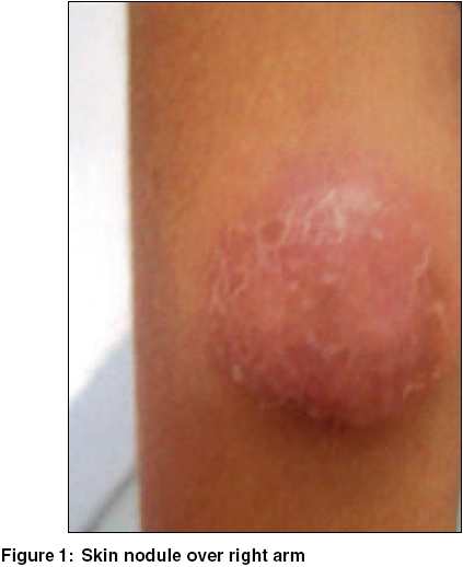
Case 1 A 14-year-old girl presented with history of painful swelling over the posterior aspect of mid-thoracic region of one-month duration. Examination revealed a tender mass measuring 7 x 8 cm in right paravertebral region at the level of D4-5. Rest of the clinical examination was unremarkable. CT scan showed a well-defined non-calcific mass abutting right paravertebral region; posterior and postero-lateral chest wall with mixed bony changes (sclerosis and lysis) in the adjacent 6th rib. FNAC from the paravertebral mass confirmed the diagnosis of Ewing′s sarcoma. Work up including bone marrow study and bone scan did not reveal any metastasis. Patient was started on induction chemotherapy comprising combination of vincristine, ifosfamide, etoposide, adriamycin and cyclophosphamide. Patient presented six weeks later with chest pain and cough. On repeat investigation her primary mass had shown considerable reduction in size but there was appearance of new lytic lesion in the 10th rib suggestive of bony metastasis. The patient was advised local radiotherapy and continuation of chemotherapy. Within the next few days the patient developed multiple skin nodules - one over right arm [Figure - 1] measuring 3 x 3 cm, one each in both supraclavicular regions measuring 3 x 3 cm and one over right lateral chest wall measuring 4 x 5 cm in size. FNAC from one of these lesions confirmed metastasis of Ewing′s sarcoma.
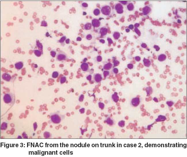
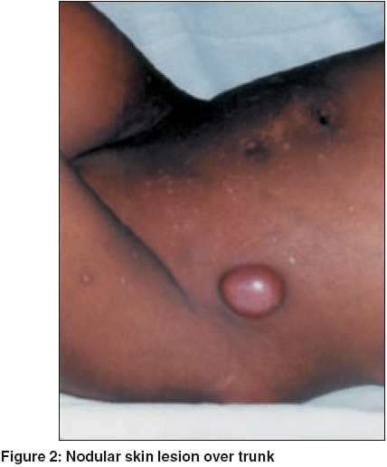
Case 2 A 7-year-old girl presented with history of pain and swelling in right gluteal region of seven months duration. She was diagnosed as Ewing′s sarcoma of right ileum with skeletal metastases (C2, D2, L3 vertebrae, right ischium and lower end of left radius) elsewhere and was treated outside with induction chemotherapy comprising vincristine, adriamycin, cyclophosphamide, ifosfamide and etoposide. She also received post induction external radiotherapy to primary site and was referred to our institute for further treatment. She presented to us with hard, tender, diffuse swelling of the right gluteal region with bilateral multiple inguinal lymphadenopathies. Rest of the examination was unremarkable. While being evaluated, she developed flesh colored multiple nodular skin lesions [Figure - 2] over trunk. These were variable in size, few of which became as large as 3-4 cm with occasional lesion showing ulceration. FNAC from the nodule on the trunk [Figure - 3] confirmed metastatic Ewing′s sarcoma. Under low power with Giemsa stain it demonstrated cellular aspirate, small clusters and singly occurring cells with minimal to moderate cytoplasm and highly mitotically active cellular morphology. She succumbed to her illness two weeks later.
Case 3 A 26 year old male symptomatic for 1 year with painful swelling of left gluteal region was diagnosed as Ewing′s sarcoma of left ileum and treated elsewhere with local radiotherapy followed by chemotherapy comprising vincristine, adriamycin, cyclophosphamide and dactinomycin. He presented to us with pain at the primary site and subcutaneous nodules over left gluteal region (not along the biopsy tract). There was no apparent swelling at the primary site. On investigation, MRI showed a large lobulated mass measuring 14 cm x 12 cm x 10 cm involving left iliac bone with intrapelvic extension. FNAC of the subcutaneous nodule was reported as metastatic (Ewing′s sarcoma) disease.
Discussion
Ewing′s family of tumors includes Ewing′s sarcoma (ES) and primitive neuro-ectodermal tumors (PNETs). They are small round cell undifferentiated neoplasm, arising from flat bones and diaphyseal regions of long bones. It is commonly found in first two decades of life. ES of chest wall is also known as Askin Rosai tumor. ES/PNET presents clinically with swelling and pain at the site of tumor. Radiologically, lytic or mixed lytic and sclerotic lesions are often seen with a soft tissue component. Histopathologically, it comprises sheets of small round cells with scanty cytoplasm and vesicular nuclei. Common sites of metastasis are lung, pleura and other bones. Less commonly lymph nodes, bone marrow, central nervous system and liver may also be involved.[1]
Cutaneous metastasis is rare. It occurs in up to 9% of all patients with cancer.[2] This may be the first sign of visceral malignancy or may develop at recurrence. They appear as non-specific painless dermal or subcutaneous nodules with intact overlying epidermis. These may be flesh colored, pink violaceous or brownish black. These are often stony hard on palpation and may become necrotic or ulcerated. Growth pattern of cutaneous metastases is unpredictable and may not reflect that of primary tumor. They have even been documented to occur within radiation sites. Depending upon the mode of spread, localization of these may differ. Tumors that tend to invade veins often present as distant cutaneous metastases.
In a thirty-year study at St. Jude′s children research hospital, Memphis, out of 197 cases of non-hematological malignancies, only 34 cases were reported to have developed cutaneous metastases of which two were of Ewing′s sarcoma.[3] Similarly Izquierdo et al reported two cases with cutaneous metastases from Ewing′s sarcoma.[4] As with any other cancer, metastasis to skin indicates a poor prognosis. We are reporting three cases of Ewing′s sarcoma who developed cutaneous metastases, which is extremely uncommon.
| 1. |
Cangir A, Vietti TJ, Gehan EA, Burgert EO Jr, Thomas P, Tefft M, et al. Ewing's sarcoma metastatic at diagnosis. Results and comparisons of two intergroup Ewing's sarcoma studies. Cancer 1990;66:887-93.
[Google Scholar]
|
| 2. |
Spencer PS, Helm TN. Skin metastases in cancer patients. Cutis 1987;39:119-21.
[Google Scholar]
|
| 3. |
Wesche WA, Khare VK, Chesney TM, Jenkins JJ. Non-hematopoetic cutaneous metastases in children and adolescent. J Cutan Path 2000;27:485-92.
[Google Scholar]
|
| 4. |
Izquierdo MJ, Sashi MA, Carrasco L, Requena C, Farina MC, Martin L, et al. Cutaneous metastases from Ewing's Sarcoma: report of two cases. Clin Exp Dermatol 2002;27:123-8.
[Google Scholar]
|
Fulltext Views
4,670
PDF downloads
3,194


