Translate this page into:
Extraskeletal chondroma of the scalp: An atypical location
Correspondence Address:
Seung-Ho Lee
Department of Dermatology, Dongguk University Ilsan Hospital, College of Medicine, Dongguk University, 814 Siksa Dong, Ilsandong gu, Goyang si, Gyeonggi do
South Korea
| How to cite this article: Choi Y, Lim WS, Lee AY, Lee SH. Extraskeletal chondroma of the scalp: An atypical location. Indian J Dermatol Venereol Leprol 2013;79:435-436 |
Sir,
As an indolent solitary subcutaneous nodule, extraskeletal chondroma usually develops in the hands and feet, and more rarely in the head and neck region of adults. [1],[2] Less than 40 cases of extraskeletal chondroma that occurred in the head and neck region have been reported worldwide. [2],[3] According to a previous study, nasal cavity, paranasal sinuses, larynx and tongue were the most reported sites in the head and neck region. [2] It has not been reported to have occurred in the scalp. We report a case of extraskeletal chondroma that occurred in the scalp, an atypical location.
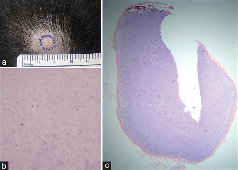
A 38-year-old Korean female presented with a palpable nodule in the scalp that had been slowly growing since it was first recognized a few months ago. The lesion had not been treated or examined until she visited us. Skin examination revealed a palpable, flesh and firm subcutaneous nodule measuring 1 × 1 cm in diameter in the frontal scalp [Figure - 1]a. The nodule was movable over the underlying bone. The patient complained of no symptoms, and tenderness was not found. Physical examination was not remarkable other than above-described skin lesion, and she had no specific past or family history.
 |
| Figure 1: (a) A palpable firm subcutaneous nodule in the scalp, measuring 1 × 1 cm in diameter. (b) An encapsulated nodule entirely composed of mature hyaline cartilage (H and E, × 20). (c) Tumor consists of oval to round chondrocytes with oblong nuclei of various size. There were no atypia or mitosis (H and E, × 100) |
Extraskeletal chondroma is a rare, benign cartilaginous tumor of the soft tissue. It presents as a solitary subcutaneous nodule or mass measuring less than 3 cm in diameter that is usually painless and slowly growing. [1] It is most frequently found in the hands and feet of adults in the fourth and fifth decades. [1] The majority has no symptoms, but Chung and Enzinger [1] reported that 19% of patients had pain and tenderness. Although the etiology of soft tissue chondroma is not completely known, it is believed that they arise from residual embryonic tissue or from uncommitted mesenchymal stem cells by either metaplastic or neoplastic process. [2]
Histologically it shows an encapsulated lobular nodule composed of mature hyaline cartilage, and is not associated with underlying bone tissues. [1] However, it may show foci of dystrophic calcification or metaplastic ossification. Occasionally, nuclear atypia, pleomorphism, binucleated lacunae, or stromal myxoid degeneration are found. Our case revealed an encapsulated nodule of mature cartilage with no cytologic atypia or mitosis. Although it can be easily diagnosed based on its typical histological findings, tumoral calcinosis, giant cell tumor, chondroblastoma and chondrosarcoma are included in the differential diagnosis. Tumoral calcinosis should be differentiated when calcification in the tumor is prominent. Cartilaginous tissues are not present and foreign body reaction is associated with tumoral calcinosis. [4] Giant cell tumor may be considered when granulomatous change is present, but it does not contain chondrocytes in the tumor. [4] Chondroblastoma should be differentiated when tumor cells resemble chondroblasts that were not found in our case. [5] Chondrosarcoma presents mitosis, atypism, and necrosis that are not seen in extraskeletal chondroma. [1] Our case was diagnosed as extraskeletal chondroma based on its typical histological pattern without atypia, mitosis or prominent stromal degeneration.
Complete excision is recommended for the treatment of extraskeletal chondroma. It is generally considered that extraskeletal chondroma is not likely to show malignant transformation, [4] because malignant change is extremely rare. We have experienced an interesting case of extraskeletal chondroma that occurred in an atypical site. To the best of our knowledge, this is the first case report that was found in the scalp.
| 1. |
Chung EB, Enzinger FM. Chondroma of soft parts. Cancer 1978;41:1414-24.
[Google Scholar]
|
| 2. |
Falleti J, De Cecio R, Mentone A, Lamberti V, Friscia M, De Biasi S, et al. Extraskeletal chondroma of the masseter muscle: A case report with review of the literature. Int J Oral Maxillofac Surg 2009;38:895-9.
[Google Scholar]
|
| 3. |
Kwon H, Kim HY, Jung SN, Shon WI, Yoo G. Extraskeletal chondroma in the auricle. J Craniofac Surg 2010;21:1990-1.
[Google Scholar]
|
| 4. |
Weiss SW, Goldblum JR. Cartilaginous soft tissue tumors. In: Weiss SW, Goldblum JR, Enzinger FM, editors. Enzinger and Weiss's soft tissue tumors. 4 th ed. St Louis: Mosby; 2001. p. 1361-8.
th ed. St Louis: Mosby; 2001. p. 1361-8.'>[Google Scholar]
|
| 5. |
Lee JW, Kim DS, Kim MK, Suh YL. Chondroblastoma-like extraskeletal chondroma. Korean J Pathol 1999;33:55-8.
[Google Scholar]
|
Fulltext Views
2,791
PDF downloads
1,372


