Translate this page into:
Extrusion of sebaceous gland into a blister of pemphigus vulgaris: An unusual processing artifact
Correspondence Address:
Rajiv Joshi
14, Jay Mahal, 'A' Road, Churchgate, Mumbai - 400 020
India
| How to cite this article: Joshi R, Marwah H S. Extrusion of sebaceous gland into a blister of pemphigus vulgaris: An unusual processing artifact. Indian J Dermatol Venereol Leprol 2004;70:316-317 |
 |
 |
Sir,
Extrusion of sebaceous glands through the follicular canal onto the skin surface is a well known phenomenon first described by Pinkus and Mehrgan.[1] Transfollicular sebaceous gland extrusion is explained as an artifact caused by damage to the fragile sebaceous gland by the effects of the physico-chemical changes that occur during tissue processing and the squeezing effect of the microtome knife as it slices through the paraffin block containing the biopsy tissue, pushing up the dislodged sebaceous gland outward through the common folliculo-sebaceous conduit.
Some authors have expressed reservations about it being an artifact, proposing instead that it be considered a natural phenomenon.[2] Sebaceous glands are found either totally extruded onto the skin surface, or may be seen in a subcorneal location.
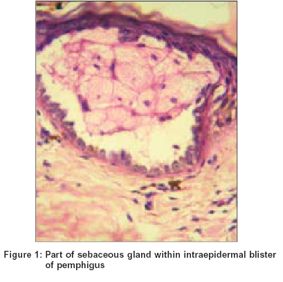
We report an unusual location of sebaceous gland extrusion, namely, within the suprabasal blister of pemphigus vulgaris.
A 34 year old male presented to one of us (HSM), with a one year history of multiple fluid filled blisters all over the body. He had earlier received short courses of oral steroids in the dose of about 15-20 mg prednisolone per day with partial clearing of the blisters. However, on discontinuing the steroids, he had developed extensive relapse with numerous small flaccid vesicles appearing on normal looking skin. Oral lesions had been present on and off, but no oral involvement was present at the time of this presentation. A clinical diagnosis of pemphigus was considered and a biopsy that included one of the new vesicles was obtained from the right side of the upper back.
Sections from the biopsy showed a small suprabasal blister with an intact sebaceous lobule occupying the cavity of the blister. [Figure - 1] This structure was identified as a sebaceous lobule as it was made up of typical mature sebocytes with central scalloped nuclei and abundant vacuolated cytoplasm. A few acantholytic cells were seen in the blister, but no inflammation or other blisters were seen in the section. Serial sections taken through the block revealed in the mid dermis a folliculo-sebaceous structure that showed suprabasal clefts with acantholysis in follicular and sebaceous epithelium, findings consistent with pemphigus vulgaris.
In sum, we report an unusual histopathological finding of extrusion of sebaceous gland into the blister cavity of pemphigus vulgaris. The acantholytic process in the lesion biopsied in our patient appears to affect the folliculo-sebaceous epithelium leading to sebaceous gland extrusion into an acantholytic suprabasal blister within the epidermis.
| 1. |
Mehregan AH, Alberta E, Pinkus H. Artifacts in dermatohistopathology. Arch Dermatol 1966;94:218-25.
[Google Scholar]
|
| 2. |
Weigand DA. Transfollicular extrusion of sebaceous glands: Natural phenomenon or artifact? A case report. J Cutan Pathol 1976;3:239-44.
[Google Scholar]
|
Fulltext Views
2,387
PDF downloads
1,040


