Translate this page into:
Finger like growth and multiple nodules over right upper back
Correspondence Address:
Mohan H Kudur
Department of Dermatology, Kasturba Medical College, Manipal-576 104, Udupi Dist, Karnataka
India
| How to cite this article: Kudur MH, Nayak S, Pai SB, Sripathi H, Prabhu S. Finger like growth and multiple nodules over right upper back. Indian J Dermatol Venereol Leprol 2010;76:589-590 |
Case History
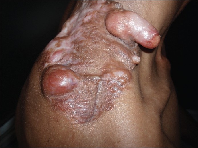
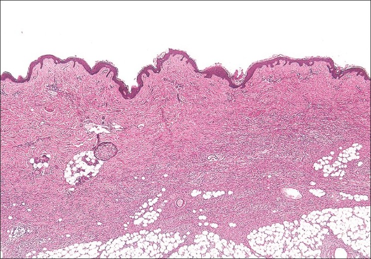
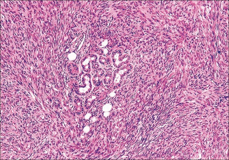
A 31-year-old male agriculturist, presented to skin OPD with asymptomatic multiple nodules over right upper back of 4 years duration. The lesion started as a single skin colored nodule over right upper back which progressed to form multiple nodules within a span of 1 year. These nodules were excised in a local hospital. After 6 months, these nodules reappeared in the same site. There is no history of trauma or any systemic illness before the onset of lesions. Examination revealed large area of atrophied skin over right upper back with multiple skin-colored nodules of varying sizes from 0.5 cm to 5 cm and a finger like growth of 6 cm [Figure - 1]. The posterior part of the lesion showed areas of depigmentation while anterior part showing hyperpigmentation. An ulcer was seen at the tip of the finger like growth. The surface of a large nodule showed telangiectasia. A skin biopsy from a small nodule showed typical histopathological features [Figure - 2] and [Figure - 3].
 |
| Figure 1 : Finger-like growth and multiple nodules seen over upper back |
 |
| Figure 2 : Epidermis showing orthokeratosis, mild acanthosis, and effacement of rete ridges. There is diffuse infiltration of spindle cells throughout dermis extending in to subcutaneous fat (H and E, ×40). |
 |
| Figure 3 : Storiform pattern of arrangement of spindle cells in dermis (H and E, ×200) |
What is Your Diagnosis?
| 1. |
Taylor HB, Helwig EB. Dermatofibrosarcoma protuberans. A study of 115 cases. Cancer 1962;15:717-25.
[Google Scholar]
|
| 2. |
Weinstein JM, Drolet BA, Esterly NB, Rogers M, Bauer BS, Wagner AM, et al. Congenital dermatofibrosarcoma protuberans: variability in presentation. Arch Dermatol 2003;139:207-11.
[Google Scholar]
|
| 3. |
Grenn JJ, Heymann WR. Dermatofibrosarcoma protuberans occurring in a small pox vaccination scar. J Am Acad Dermatol 2003;48:S54-55.
[Google Scholar]
|
| 4. |
Parlette LE, Smith CK, Germain LM, Rolfe CA, Skelton H. Accelerated growth of dermatofibrosarcoma protuberans during pregnancy. J Am Acad Dermatol 1999;41:778-83.
[Google Scholar]
|
| 5. |
Resnik KS, DiLeonardo M, Hunter CJ. Pedunculated presentation of dermatofibrosarcoma protuberans. J Am Acad Dermatol 2003;49:1139-41.
[Google Scholar]
|
| 6. |
Gφkden N, Dehner LP, Zhu X, Pfeifer JD. Dermatofibrosarcoma protuberans of the vulva and groin: detection of COL1A1 - PDGFB fusion transcripts by RT-PCR. J Cutan Pathol 2003;30:190-5.
[Google Scholar]
|
| 7. |
Chang CK, Jacobs IA, Salti GI. Outcomes of surgery for dermatofibrosarcoma protuberans. Eur J Surg Oncol 2004;30:341-5.
[Google Scholar]
|
| 8. |
Aiba S, Tabata N, Ishii H, Ootani H, Tagami H. Dermatofibrosarcoma protuberans is a unique fibrihistiocytic tumor expressing CD34. Br J Dermatol 1992;172:79-84.
[Google Scholar]
|
Fulltext Views
2,374
PDF downloads
1,790


