Translate this page into:
Focal dermal hypoplasia (Goltz syndrome)
2 Departments of Pediatrics, Medical College, Calicut, Kerala, India
Correspondence Address:
Najeeba Riyaz
"Arakkal", Chalappuram Calicut 2, Kerala- 673 002
India
| How to cite this article: Riyaz N, Riyaz A, Chandran R, Rakesh S V. Focal dermal hypoplasia (Goltz syndrome). Indian J Dermatol Venereol Leprol 2005;71:279-281 |
Abstract
A 7-year-old girl born of non-consanguineous marriage was evaluated for facial dysmorphism. She had multiple skeletal anomalies like hypoplasia of the right mandible, narrow nasal bridge with broad tip and unilateral notching of the right ala nasi, concomitant squint and low set ears. She also had generalized hypopigmented, atrophic linear macules, multiple papillomas, fat herniations, umbilical hernia, hypoplastic nails, cicatricial alopecia, mild mental retardation, 'lobster-claw' hand and osteopathia striata of long bones, pointing to a diagnosis of Goltz syndrome. The unusual features noted were absence of the left first rib and aortic regurgitation. |
 |
 |
 |
INTRODUCTION
Focal dermal hypoplasia (FDH) is a rare mesoectodermal disorder inherited by an X-linked dominant gene, which is lethal in homozygous males. Occasional occurrence in males is due to fresh mutations. It was first described by Goltz in 1962.[1] Of the 175 cases reported worldwide, there are only three from India.[2],[3],[4]
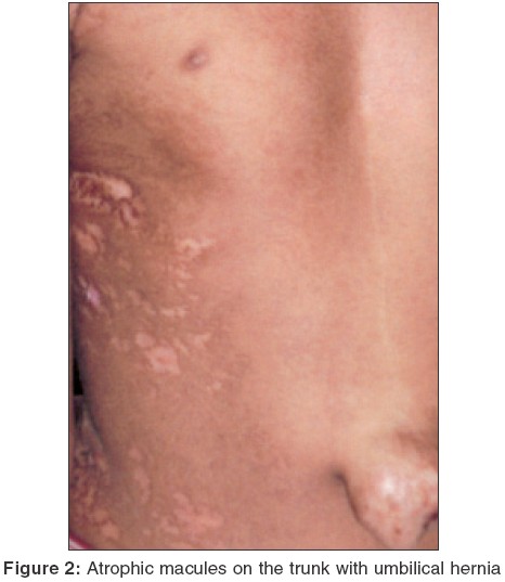
A 7-year-old girl, born of non-consanguineous parents, was evaluated for facial dysmorphism [Figure - 1]. She was born by normal vaginal delivery. She weighed 2.5 kg at birth and had multiple skeletal anomalies, facial dysmorphism and hypopigmented skin lesions over the trunk and extremities at the time of birth. All her milestones of development were slightly delayed. Her 10-year-old brother was normal. Her mother had history of two abortions with gestational age of 2 months each.
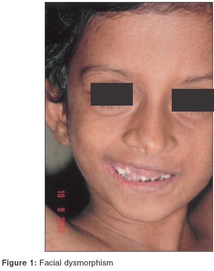
On examination, her height was 91 cm (expected 120 cm) showing Grade IV stunting. Her weight was 18 kg, the expected being 22 kg. She had facial asymmetry. The right mandible was hypoplastic and the nasal bridge was narrow with a broad tip and unilateral notching of right ala nasi. She had concomitant squint and low set ears. Her teeth were hypoplastic and maloccluded with enamel hypoplasia. The lower incisors were notched. Her right hand was split with syndactyly and the middle finger was absent (lobster-claw hand). The right first and second toes were fused and the third toe was absent. She also had scoliosis and an umbilical hernia [Figure - 2]. Her IQ was around 60-70, indicative of mild mental retardation.
Cutaneous examination showed multiple atrophic, pigmented, linear streaks and macules of varying sizes on the trunk and extremities along the lines of Blaschko, resembling striae. Multiple papillomatous and angiofibromatous lesions were seen at the right angle of the mouth and at the perianal region. Soft yellowish, lipomatous nodules projecting through localized areas of skin atrophy were seen on the legs. Multiple patchy areas of cicatricial alopecia were seen on the vertex. Nails were narrow, hypoplastic and dystrophic.
Systemic examination was essentially normal but echocardiogram showed mild aortic regurgitation.
Her routine hemogram and urine analysis were normal. X-rays of long bones showed the characteristic ′osteopathia striata′ (longitudinal striations in metaphysis of long bones). X-rays of hands and feet showed hypoplastic metacarpals, syndactyly and absent right middle finger. Chest X-ray showed absence of the left first rib. Ultrasound scan of abdomen did not reveal any abnormality. Skin biopsy showed marked dermal atrophy with thin collagen fibers consistent with focal dermal hypoplasia.
DISCUSSION
Goltz syndrome has a multitude of clinical features. The characteristic features include atrophic linear hypo- or hyperpigmented patches, fat herniation through dermal defects, and multiple papillomas of the mucous membranes or skin. Dental anomalies include hypodontia, oligodontia, microdontia, enamel fragility and dysplasia, retarded eruption and malocclusion.[3] Warburg observed microphthalmia with bilateral coloboma of the iris and ectopia lentis.[5] Other ocular lesions described are strabismus and anophthalmia.
Skeletal anomalies include asymmetric involvement of the hands and feet in 60% of patients, including syndactyly, ectrodactyly, polydactyly, absence or hypoplasia of digits and even absence of an extremity. Cervical rib has been reported.[6] Scoliosis occurs in 20% of cases. Skeletal asymmetry, clavicular dysplasia and spina bifida occulta can occur. The characteristic radiological change is osteopathia striata of the long bones.[7] Our patient had multiple skeletal anomalies like osteopathia striata, hypoplastic metacarpals, syndactyly and absent right middle finger. Also, her left first rib was absent.
Goltz reviewed this disorder in 1992.[8] Patients with areas of total absence of skin at birth have been reported. Apocrine gland anomalies and hidrocystomas near the eyes also have been described. Fibrovascular papillomas, especially in the perianal and vulvar regions, are sometimes mistaken for condylomas. Rarely, laryngeal and esophageal papillomas are seen. Osteopathia striata is a frequent finding and ′lobster-claw′ hand is a striking feature of Goltz syndrome. Occasional anomalies include short stature, joint hypermobility, mental retardation (15%), hearing defects, microcephaly, horse-shoe kidneys, umbilical, inguinal, epigastric, or diaphragmatic hernias.[9] Cardiac anomalies include cardiac tumors[10] and congenital heart diseases like truncus arteriosus.[11] Of all these features, our patient had umbilical hernia and mental retardation.
The vast majority of cases of FDH are sporadic and the affected persons are females. Recently it has been postulated that the gene is located at the Xp terminal region.[12] Our patient had all the typical features of Goltz syndrome. The additional features present in this case were absence of the left first rib and mild aortic regurgitation.
| 1. |
Goltz RW, Peterson WC Jr, Gorlin RJ, Ravits HG. Focal dermal hypoplasia. Arch Derm 1962;86:708-17.
[Google Scholar]
|
| 2. |
Sule RR, Dhumawat DJ, Gharpuray MB. Focal dermal hypoplasia. Cutis 1994;53:309-12.
[Google Scholar]
|
| 3. |
Premalatha S, Augustine SM, Thambaiah AS. Focal dermal hypoplasia syndrome - a case report. Indian Pediatr 1978:15:443-4.
[Google Scholar]
|
| 4. |
Premalatha S, Kalyani NM, Janaki VR, Chandrasekharan A, Thambaiah AS. Focal dermal hypoplasia syndrome: a case report. Indian J Pediatr 1981;48:781-3.
[Google Scholar]
|
| 5. |
Warburg M. Focal dermal hypoplasia: ocular and general manifestations with a survey of the literature. Acta Ophthal 1970;48:525-36.
[Google Scholar]
|
| 6. |
Ogunbiyi AO, Adewole IO, Ogunleye O, Ogunbiyi JO, Ogunseinde OO, Baiyeroju-Agbeja A. Focal dermal hypoplasia: a case report and review of literature. West Afr J Med 2003;22:346-9.
[Google Scholar]
|
| 7. |
Goltz RW, Henderson RR, Hitch JM, Ott JE. Focal dermal hypoplasia syndrome. A review of the literature and report of two cases. Arch Derm 1970;101:1-11.
[Google Scholar]
|
| 8. |
Goltz RW. Focal dermal hypoplasia syndrome: An update. Arch Derm 1992;128:1108-11.
[Google Scholar]
|
| 9. |
Patel JS, Maher ER, Charles AK. Focal dermal hypoplasia Goltz syndrome presenting as a severe fetal malformation syndrome. Clin Dysmorphol 1997;6:267-72.
[Google Scholar]
|
| 10. |
Doede T, Seidel J, Riede FT, Vogt L, Mohr FW, Schier F. Occult, life threatening, cardiac tumor in syndactylism in Gorlin-Goltz syndrome. J Pediatr Surg 2004;39:e17-9.
[Google Scholar]
|
| 11. |
Han XY, Wu SS, Conway DH, Pawel BR, Punnett HH, Martin RA. Truncus arteriosus and other lethal internal anomalies in Goltz syndrome. Am J Med Genet 2000;90:45-8.
[Google Scholar]
|
| 12. |
Naritomi K, Izumikawa Y, Nagataki S, Fukushima Y, Wakui K, Niikawa N, et al. Combined Goltz and Alicardi syndromes in a terminal XP deletion; are they a contiguous gene syndrome? Am J Med Genet 1992;43:839-43.
[Google Scholar]
|
Fulltext Views
4,567
PDF downloads
3,096


