Translate this page into:
Follicular lichen planus pigmentosus in blaschkoid pattern: Superimposed segmental mosaicism
2 Department of Histopathology, Postgraduate Institute of Medical Education and Research, Chandigarh, India
Correspondence Address:
Muthu Sendhil Kumaran
Department of Dermatology, Venereology and Leprology, Postgraduate Institute of Medical Education and Research, Sector 12, Chandigarh - 160 012
India
| How to cite this article: Daroach M, Guliani A, Keshavmurthy V, Vishwajeet V, Saikia UN, Kumaran MS. Follicular lichen planus pigmentosus in blaschkoid pattern: Superimposed segmental mosaicism. Indian J Dermatol Venereol Leprol 2020;86:305-307 |
Sir,
Lichen planus pigmentosus presents as slate-grey pigmented macules and patches predominantly affecting the head and neck. Follicular lichen planus pigmentosus is a rare variant characterized by greyish follicular macules.[1] Only few cases have been reported so far. Here, we present a case of follicular lichen planus pigmentosus in a blaschkoid pattern and briefly discuss genetic mosaicism.
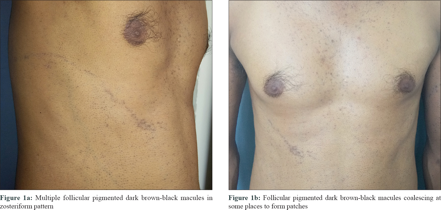
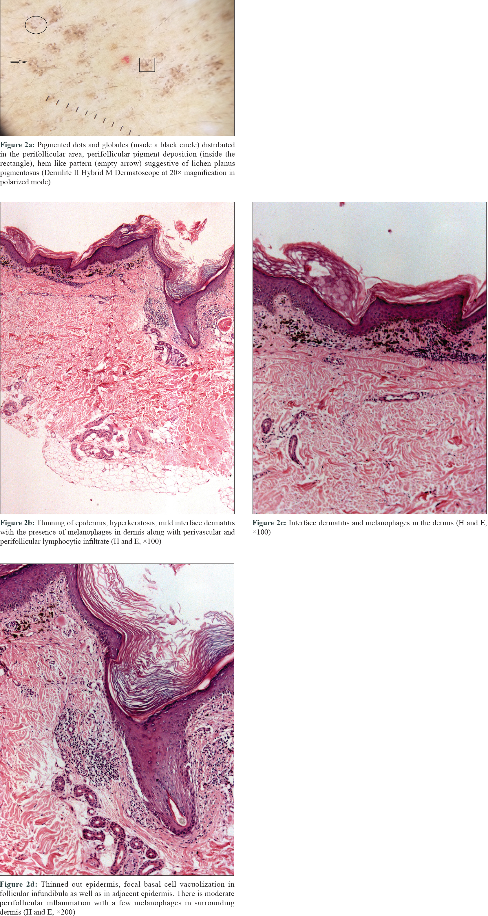
A 28-year-old man presented to our outpatient department with a history of asymptomatic pigmented patches on the face, neck, chest and abdomen from the last 2 years. The pigmentation initially appeared on the face and neck and later progressed to involve the right infrascapular area and right side of the abdomen in a linear pattern over 5 months. There was no history of any drug intake, trauma prior to the eruption and occurrence of herpes zoster. On cutaneous examination, multiple linear, follicular, pigmented macules were present on the right side of the abdomen extending to the back in a blaschkoid pattern [Figure - 1]a. These macules were 0.2 to 0.6 cm in size, irregular in shape and were well circumscribed. Some of the lesions showed minimal atrophy. Multiple follicular hyperpigmented macules coalescing at some places to form patches were seen on the face, neck and chest [Figure - 1]b. Examination of oral cavity, genitals, scalp and nails was normal. Dermoscopy revealed pigment dots and globules, “hem-like” pattern and pigmentation in the perifollicular area suggestive of lichen planus pigmentosus [Figure - 2]a. Histopathological examination of a hyperpigmented macule revealed focal thinned out epidermis. There was patchy basal cell vacuolization in follicular infundibula as well as in adjacent epidermis and moderate perifollicular inflammation with melanophages in the surrounding dermis, compatible with the diagnosis of lichen planus pigmentosus [Figure - 2]b, [Figure - 2]c, [Figure - 2]d. He was prescribed topical tacrolimus 0.1% ointment once a day.
 |
| Figure 1: |
 |
| Figure 2: |
Lichen planus pigmentosus is not an uncommon pigmentary disorder in Asian populations.[1] It can present as diffuse, reticular, blotchy, perifollicular patterns and rarely as linear, zosteriform and inverse patterns. Follicular lichen planus pigmentosus is a recently described variant, thought to develop during disease exacerbation, commonly over the trunk and upper limb, with a female preponderance.[1] In this case, follicular lichen planus pigmentosus lesions started from the face and later involved the trunk and extremities. Lesions were differentiated from lichen plano pilaris and follicular lichen planus on basis of dermatoscopic patterns (pigment dots and globules, “hem-like” pattern and perifollicular pigmentation) and histopathological features.[2] Interestingly, the present case developed blaschkoid lesions over abdominal wall and back superimposed on generalized manifestation, which might represent 'superimposed segmental manifestation,' a type of genetic mosaicism described in polygenic inflammatory disorders like lichen planus, psoriasis, vitiligo, pemphigus vulgaris, granuloma annulare, dermatomyositis and systemic lupus erythematosus.[3] Lines of blaschko are postulated to correspond to the migration of ectodermal and neuroectodermal cells during embryogenesis and also thought to reflect T-lymphocytic migration and clonal expression during embryogenesis of skin.[4] A loss of heterozygosity of one of the concerned genes distributed along lines of Blaschko leads to superimposed segmental manifestation.[3] Linear lesions are usually seen in lichen planus; however, there are only a few reports of blaschkoid lichen planus pigmentosus.[5],[6] We were unable to find a previous similar report of blaschkoid follicular lichen planus pigmentosus in literature. Future research should focus on the genetics of lichen planus pigmentosus and the role of T cells in disease pathogenesis.
Declaration of patient consent
The authors certify that they have obtained all appropriate patient consent forms. In the form, the patient has given his consent for his images and other clinical information to be reported in the journal. The patient understands that name and initials will not be published and due efforts will be made to conceal the identity, but anonymity cannot be guaranteed.
Financial support and sponsorship
Nil.
Conflicts of interest
There are no conflicts of interest.
| 1. |
Sindhura KB, Vinay K, Kumaran MS, Saikia UN, Parsad D. Lichen planus pigmentosus: A retrospective clinico-epidemiologic study with emphasis on the rare follicular variant. J Eur Acad Dermatol Venereol 2016;30:e142-4.
[Google Scholar]
|
| 2. |
Vinay K, Bishnoi A, Parsad D, Saikia UN, Sendhil Kumaran M. Dermatoscopic evaluation and histopathological correlation of acquired dermal macular hyperpigmentation. Int J Dermatol 2017;56:1395-9.
[Google Scholar]
|
| 3. |
Kouzak SS, Mendes MS, Costa IM. Cutaneous mosaicisms: Concepts, patterns and classifications. An Bras Dermatol 2013;88:507-17.
[Google Scholar]
|
| 4. |
Seo JK, Lee HJ, Lee D, Choi JH, Sung HS. A case of linear lichen planus pigmentosus. Ann Dermatol 2010;22:323-5.
[Google Scholar]
|
| 5. |
Vineet R, Sumit S, K GV, Nita K. Lichen planus pigmentosus in linear and zosteriform pattern along the lines of Blaschko. Dermatol Online J 2015;21. pii: 13030/qt4rk2w3rm.
[Google Scholar]
|
| 6. |
Akagi A, Ohnishi Y, Tajima S, Ishibashi A. Linear hyperpigmentation with extensive epidermal apoptosis: A variant of linear lichen planus pigmentosus? J Am Acad Dermatol 2004;50:S78-80.
[Google Scholar]
|
Fulltext Views
5,746
PDF downloads
3,075


