Generic selectors
Exact matches only
Search in title
Search in content
Post Type Selectors
Search in posts
Search in pages
Filter by Categories
15th National Conference of the IAOMFP, Chennai, 2006
Abstract
Abstracts from current literature
Acne in India: Guidelines for management - IAA Consensus Document
Addendum
Announcement
Art & Psychiatry
Article
Articles
Association Activities
Association Notes
Award Article
Book Review
Brief Report
Case Analysis
Case Letter
Case Letters
Case Notes
Case Report
Case Reports
Clinical and Laboratory Investigations
Clinical Article
Clinical Studies
Clinical Study
Commentary
Conference Oration
Conference Summary
Continuing Medical Education
Correspondence
Corrigendum
Cosmetic Dermatology
Cosmetology
Current Best Evidence
Current Issue
Current View
Derma Quest
Dermato Surgery
Dermatopathology
Dermatosurgery Specials
Dispensing Pearl
Do you know?
Drug Dialogues
e-IJDVL
Editor Speaks
Editorial
Editorial Remarks
Editorial Report
Editorial Report - 2007
Editorial report for 2004-2005
Errata
Erratum
Focus
Fourth All India Conference Programme
From Our Book Shelf
From the Desk of Chief Editor
General
Get Set for Net
Get set for the net
Guest Article
Guest Editorial
History
How I Manage?
IADVL Announcement
IADVL Announcements
IJDVL Awards
IJDVL AWARDS 2015
IJDVL Awards 2018
IJDVL Awards 2019
IJDVL Awards 2020
IJDVL International Awards 2018
Images in Clinical Practice
Images in Dermatology
In Memorium
Inaugural Address
Index
Knowledge From World Contemporaries
Leprosy Section
Letter in Response to Previous Publication
Letter to Editor
Letter to the Editor
Letter to the Editor - Case Letter
Letter to the Editor - Letter in Response to Published Article
LETTER TO THE EDITOR - LETTERS IN RESPONSE TO PUBLISHED ARTICLES
Letter to the Editor - Observation Letter
Letter to the Editor - Study Letter
Letter to the Editor - Therapy Letter
Letter to the Editor: Articles in Response to Previously Published Articles
Letters in Response to Previous Publication
Letters to the Editor
Letters to the Editor - Letter in Response to Previously Published Articles
Letters to the Editor: Case Letters
Letters to the Editor: Letters in Response to Previously Published Articles
Media and news
Medicolegal Window
Messages
Miscellaneous Letter
Musings
Net Case
Net case report
Net Image
Net Images
Net Letter
Net Quiz
Net Study
New Preparations
News
News & Views
Obituary
Observation Letter
Observation Letters
Oration
Original Article
ORIGINAL CONTRIBUTION
Original Contributions
Pattern of Skin Diseases
Pearls
Pediatric Dermatology
Pediatric Rounds
Perspective
Presedential Address
Presidential Address
Presidents Remarks
Quiz
Recommendations
Regret
Report
Report of chief editor
Report of Hon : Treasurer IADVL
Report of Hon. General Secretary IADVL
Research Methdology
Research Methodology
Resident page
Resident's Page
Resident’s Page
Residents' Corner
Residents' Corner
Residents' Page
Retraction
Review
Review Article
Review Articles
Reviewers 2022
Revision Corner
Self Assessment Programme
SEMINAR
Seminar: Chronic Arsenicosis in India
Seminar: HIV Infection
Short Communication
Short Communications
Short Report
Snippets
Special Article
Specialty Interface
Studies
Study Letter
Study Letters
Supplement-Photoprotection
Supplement-Psoriasis
Symposium - Contact Dermatitis
Symposium - Lasers
Symposium - Pediatric Dermatoses
Symposium - Psoriasis
Symposium - Vesicobullous Disorders
SYMPOSIUM - VITILIGO
Symposium Aesthetic Surgery
Symposium Dermatopathology
Symposium-Hair Disorders
Symposium-Nails Part I
Symposium-Nails-Part II
Systematic Review and Meta-Analysis
Systematic Reviews and Meta-analyses
Systematic Reviews and Meta-analysis
Tables
Technology
Therapeutic Guideline-IADVL
Therapeutic Guidelines
Therapeutic Guidelines - IADVL
Therapeutics
Therapy
Therapy Letter
Therapy Letters
View Point
Viewpoint
What’s new in Dermatology
View/Download PDF

Translate this page into:

Letter to the Editor
2012:78:5;654-656
doi: 10.4103/0378-6323.100573

Generalized morphea developing in a patient previously affected with eosinophilic fasciitis

Resham J Vasani1 , Sudhir V Medhekar2
1 Department of Dermatology, Venereology and Leprosy, K J Somaiya Medical College, Sion, Mumbai, Maharashtra, India
2 Department of Dermatology, Venereology and Leprosy, Shri Bhausaheb Hire Government Medical College, Dhule, Maharashtra, India

Correspondence Address:
Resham J Vasani
A/1 Sharad Kunj, Dr. Moose Road, Thane (west)
India
How to cite this article:
Vasani RJ, Medhekar SV. Generalized morphea developing in a patient previously affected with eosinophilic fasciitis. Indian J Dermatol Venereol Leprol 2012;78:654-656
Copyright: (C)2012 Indian Journal of Dermatology, Venereology, and Leprology

Sir,

A forty-four-year-old female presented with diffuse swelling with brownish tender induration of forearms and legs for the past one month. She had no history of fever, joint pains, mucosal lesions, dysphagia, dyspnoea, Raynaud′s phenomenon or proximal muscle weakness.

The clinical examination showed minimally erythematous areas of induration distributed symmetrically over flexors of forearms and legs [Figure - 1]a. On raising the forearm, there was linear furrowing along the blood vessels suggestive of dry river bed sign. A clinical diagnosis of eosinophilic fasciitis (EF) was made.

Investigations showed the presence of peripheral eosinophilia (absolute eosinophil count -1206), raised ESR (105 mm at the end of 1 h), raised IgG levels of 2000 IU/ml (N- 700-1700 IU/ml), positive ANA (1:100 in a speckled pattern), Anti Scl 70 and Anti centromere antibodies were negative. The hepatic, renal, thyroid profiles and chest radiograph were normal. The histopathological examination showed the presence of normal epidermis with the dermis showing widening of the fascial planes and septae with predominant eosinophilic infiltrate [Figure - 1]b and c. Thus, the diagnosis of EF was established.

The patient was started on oral prednisolone at the dose of 60 mg and gradually tapered over three months with subjective and objective improvement of the skin lesions.
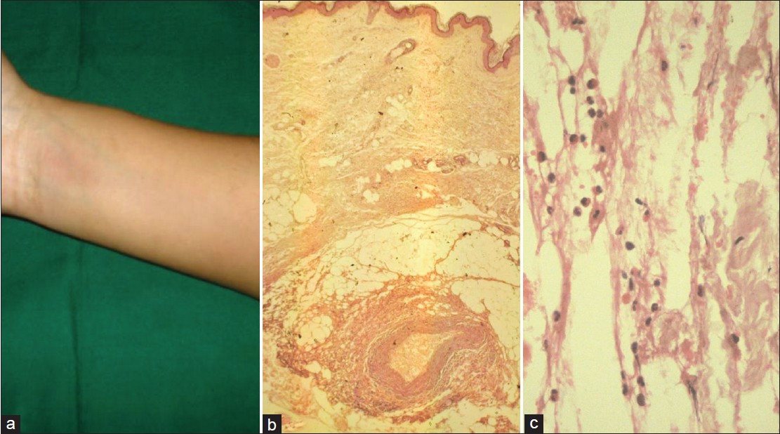
Figure 1: (a) Ill-defined area of induration with overlying skin showing mild erythema over the flexor aspect of the forearm. (b) (H and E, ×4) - Normal epidermis and dermis with thickened fascial planes showing mixed infiltrate. (c) (H and E, ×40) - Predominance of eosinophils in the fascial infiltrate

One year later, the patient presented with symptoms of multiple reddish to dark colored lesions on the trunk. The lesions were associated with itching and tightness overlying the lesions. Similar lesions were also present over the forearms and legs giving rise to restricted movements at the bilateral wrist joints.

Both general and systemic examination were normal. Dermatological examination showed the presence of multiple ill-defined, depressed, hyperpigmented plaques with thinned out; atrophic, shiny skin overlying them [Figure - 2]a. The plaques varied from 2 cm to 30 cm in their greatest dimensions. They were present over the back, chest, abdomen and proximal extremities. Rest of the cutaneous areas including the acral areas, palms, soles and mucosae were normal.
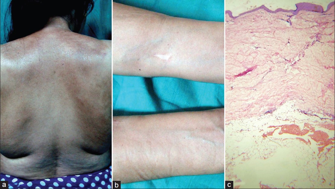
Figure 2: (a) Multiple ill-defined, depressed, hyperpigmented plaques with thinned out, atrophic, shiny skin on the back and proximal extremities of generalized morphea. (b) Previous site of eosinophilic fasciitis showing residual depression of the affected areas with prominence of the veins traversing the area. (c) (H and E, ×4) - Atrophic epidermis, thick, deeply eosinophilic, closely packed bundles of collagen in the reticular dermis and subcutis s/o morphea

The areas on the forearms and legs which showed changes of EF earlier, now showed residual depression of affected areas with prominence of veins traversing the area [Figure - 2]b. The clinical diagnosis was generalized morphea (GM).

This time investigations showed a normal eosinophil count with raised ESR (38 mm at end of 1 h), positive ANA (1:100, speckled pattern), negative anti centromere antibody and Anti Scl 70 antibody. Chest radiograph, liver, renal, thyroid profiles and abdominal ultrasound were normal. Histopathological examination showed atrophic epidermis, thick deeply eosinophilic, closely packed bundles of collagen in the reticular dermis and subcutis with atrophic appendages confirming morphea [Figure - 2]c. The patient was started on methotrexate 10 mg per week, but was lost to follow up thereafter. The final diagnosis was GM developing in an old case of EF.

It is controversial whether GM and EF are two distinct entities, whether EF is a variant of deep morphea [1] or whether EF and morphea represent a continuum of the same disease.

EF is characterized by an acute onset usually precipitated by strenuous exercise. [2] There is diffuse symmetrical involvement, with peripheral eosinophilia and hypergammaglobulinemia. The histology is specific with marked fibrosis and diffuse infiltration of the fascia with eosinophils. It is usually well responsive to oral corticosteroids.

GM usually has an insidious onset, with widespread involvement of more than two anatomic areas with well circumscribed ivory white plaques surrounded by lilac border in early stages and hyperpigmentation as lesions evolve. Poor response to corticosteroid treatment is noted.

There are certain overlapping features as follows. Both EF and morphea show similar sclerosis of skin, immunological pathogenesis, absence of sclerodactyly, visceral involvement and Raynaud′s phenomenon. Although peripheral and tissue eosinophilia and hypergammaglobulinemia are typically seen in EF, they are not the pre requisites for diagnosis [3] and can also be seen in early stages of morphea. The inflammation of the fascia in EF can extend to the adjacent dermis and also the dermal inflammation of morphea extends to the fascia as in the pansclerotic variant. Though presence of fascial inflammation and fibrosis are more consistent features of EF than morphea, it is possible that failure to routinely observe fascial fibrosis in morphea may be due to inadequate depth of biopsy which usually does not involve the subcutis. Concurrent lesions of morphea is noted in 25 to 30% cases of EF. [4] There have been case reports of transition of EF to localized GM [5] and even systemic sclerosis.

Hence to conclude, while clinical presentation in conjunction with histological details in most of the instances will allow one to differentiate EF from scleroderma, there is undeniable evidence that these two entities do share certain morphologic features that in late stages of these diseases may make their separation difficult warranting further study and long-term follow up of these patients.

References
1.
Naschitz JE, Boss JH, Misselevich I, Yeshurun D, Rosner I. The fasciitis-panniculitis syndromes: Clinical and pathologic features. Medicine (Baltimore) 1996;75:6-16.
[Google Scholar]
2.
Lakhanpal S, Ginsburg WW, Michet CJ, Doyle JA, Moore SB. Eosinophilic fasciitis: Clinical spectrum and therapeutic response in 52 cases. Semin Arthritis Rheum 1988;17:221-31.
[Google Scholar]
3.
Jensen E, Hess B, Hunziker T, Roos F, Helbling A. Eosinophilic fasciitis (Shulman syndrome). Schweiz Med Wochenschr 2000;130:156-60.
[Google Scholar]
4.
Antic M, Lautenschlager S, Itin PH. Eosinophilic fasciitis 30 years after - what do we really know? Report of 11 patients and review of the literature. Dermatology 2006;213:93-101.
[Google Scholar]
5.
Jaratt M, Bybee JD, Ramsdell W. Eosinophilic fasciitis: An early variant of scleroderma. J Am Acad Dermatol 1979;1:221-6.
[Google Scholar]

Fulltext Views
2,547

PDF downloads
1,130
Show Sections