Translate this page into:
Giant cerebriform intradermal melanocytic nevus
Corresponding author: Dr. Guang-Wen Yin, Department of Dermatology, The First Affiliated Hospital of Zhengzhou University, Zhengzhou, China. gwyin67@126.com
-
Received: ,
Accepted: ,
How to cite this article: Yin GW, Geng MM. Giant cerebriform intradermal melanocytic nevus. Indian J Dermatol Venereol Leprol 2021;87:876-7.
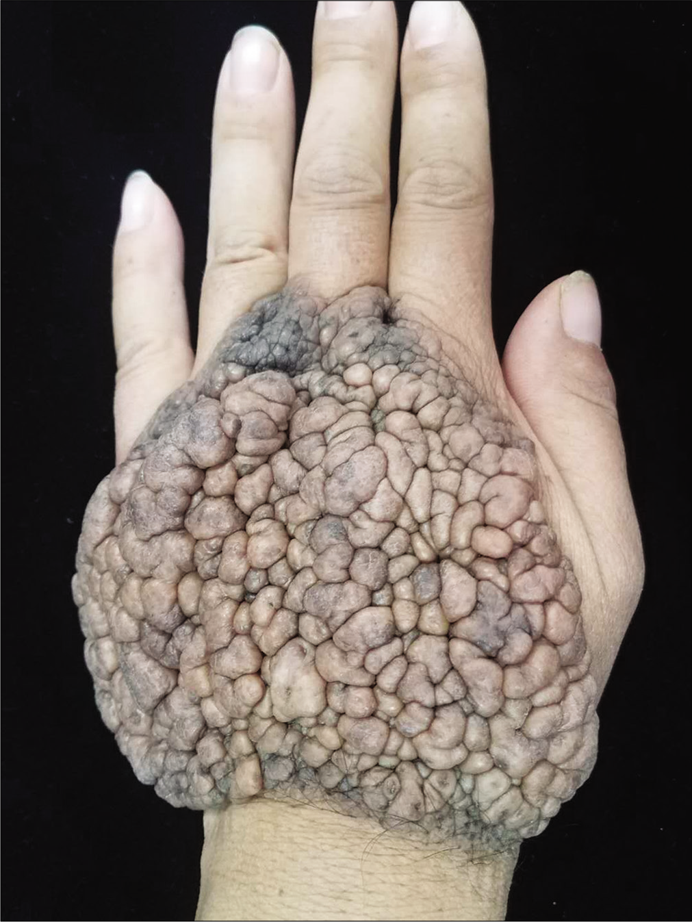
A 43-year-old woman presented with a large brownish-yellow raised lesion on the dorsal aspect of her left hand which was present since birth. Examination revealed a large cerebriform plaque measuring 11.0 × 9.0 × 3.0 cm [Figure 1], the surface of which seemed studded with hundreds of pebble-like, brownish-yellow lesions of varying sizes, with a soft and smooth texture. The remaining skin and appendages did not show any abnormality. A histopathological examination revealed solitary and variably-sized clusters of nevus cells scattered in the dermis, containing a heterogeneous amount of melanin. The patient was advised excision of the lesions.

- The large plaque on the dorsum of the left hand, surface studded with pebble-like, brownish-yellow plaques of varying sizes
Declaration of patient consent
The authors certify that they have obtained all appropriate patient consent forms. In the form the patient has given her consent for her images and other clinical information to be reported in the journal. The patient understands that her name and initials will not be published and due efforts will be made to conceal her identity but anonymity cannot be guaranteed.
Financial support and sponsorship
Nil.
Conflicts of interest
There are no conflicts of interest.


