Translate this page into:
Giant eccrine spiradenoma mimicking a malignant tumor
2 Department of Plastic, Reconstructive and Cosmetic Surgery, Apollo Hospitals, Bhubaneswar, Odisha, India
3 Department of Pathology and Oncopatholgy, Apollo Hospitals, Bhubaneswar, Odisha, India
Correspondence Address:
Satyabrata Tripathy
Consultant Dermatologist, Apollo Hospitals, Sainik School Road, Unit 15, Bhubaneswar - 751 005, Odisha
India
| How to cite this article: Tripathy S, Mishra L, Baisakh M, Mohapatra N, Das S. Giant eccrine spiradenoma mimicking a malignant tumor. Indian J Dermatol Venereol Leprol 2015;81:79-80 |
Sir,
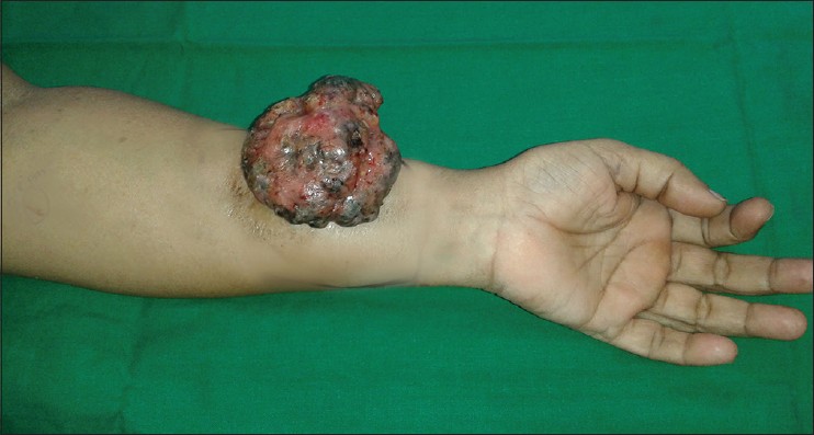
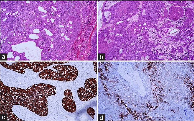
A 55-year-old female presented with a small warty protuberance on the right forearm that rapidly progressed to a large, painful growth over 6 months. Local examination revealed a cerebriform tumor of size 7 × 4.5 cm with hemorrhagic crusts on the surface [Figure - 1]. The growth was tender, mobile, not associated with regional adenopathy and bled on touch. A computed tomography (CT) scan of the affected area showed that the tumor was localized to the subcutaneous and muscular plane, without any bony involvement. Considering the size of the tumor, a surgical excision was planned and a complete excision with adequate margins was done. On histopathological examination, the lesion showed a lobulated highly cellular tumor containing a biphasic population of cells, one with dark nuclei and the other with large vesicular nuclei [[Figure - 2]a and b]. Frequent variable sized duct formation was seen surrounded by tumor cells and containing eosinophilic periodic acid-Schiff (PAS) positive material in the lumen. [[Figure - 2]a and b].The surface showed necrotic debris and polymorphs. An immunohistochemical study showed strong cytoplasmic reactivity of the tumor cells for cytokeratin (CK) antigen and S100 reactivity was seen in the secretory cells scattered between the tumor cells [[Figure - 2]c and d]. Based on these features a diagnosis of eccrine spiradenoma was made.
 |
| Figure 1: A verrucous tumor of size 7 × 4.5 cm with hemorrhagic crusts over right forearm |
 |
| Figure 2: (a) Lobules of branching nests of tumor cells are seen with areas of ductal differentiation (H and E, x20) (b) Focal areas of squamous differentiation and lymphocytic infiltration in the stroma (H and E, x20) (c) Immunohistochemistry showing strong cytoplasmic reactivity of the tumor cells toCK (IHC, CK, x40) (d) Immunohistochemistry showing S100 reactivity of the secretory cells interspersed between the tumor cells. (IHC, S100, x40) |
Eccrine spiradenoma is a rare benign tumor of sweat gland origin which is distinguished by its characteristic histology. [1] The classical presentation is a solitary, firm, painful, rounded, bluish dermal nodule, 3-50 mm in diameter, affecting the trunk or proximal extremities in a young adult. [1],[2] Rare sites include vulva, breast, external ears, nail folds and face. [1] Multiple and multifocal lesions have also been rarely described in zosteriform, linear and Blaschkoid distribution. [1],[2],[3] Multiple eccrine spiradenomas can be a part of Brooke-Spiegler syndrome, an autosomal dominant condition, with numerous tumors of various types like cylindromas, and trichoepitheliomas. [1]
The clinical differential diagnoses include hidradenoma, chondroid syringoma, leiomyoma, neuroma, angiolipoma, trichilemmal cyst and glomus tumor. Biopsy is essential to arrive at the correct diagnosis. The case described herein is unusual as it differed from the classical presentation of an eccrine spiradenoma in having a giant size and verrucous rough hemorrhagic surface. In fact, this appearance closely mimicked malignant lesions like nodular melanoma, angiosarcoma or squamous cell carcinoma. It was only after biopsy and histopathologic study that the diagnosis of eccrine spiradenoma was reached. Giant vascular eccrine spiradenoma is another variant of eccrine spiradenoma which was considered in the differential diagnosis, as it shows increased vascularity and rapid growth, reaching an abnormally large size. [4] However our case did not reveal increased vascularity on histopathology. Another close differential diagnosis is nodular hidradenoma/clear cell hidradenoma. However, presence of biphasic population of cells with frequent duct formation and absence of clear cell changes excluded this diagnosis. Malignant transformation to eccrine spiradenoma is extremely rare and is characterized by features such as rapid increase in size, bleeding, change in color, and ulceration. [1],[4],[5] Our case showed most of the clinical features of a malignant lesion but showed benign histologic findings. The patient has completed one year of follow up without any local recurrence.
| 1. |
Calonje E. Tumours of the Skin Appendages. In: Burns T, Breathnach S, Cox N, Griffiths C, editors. Rook's Textbook of Dermatology. 8 th ed. Oxford: Wiley-Blackwell; 2010. p. 53.29-30.
th ed. Oxford: Wiley-Blackwell; 2010. p. 53.29-30.'>[Google Scholar]
|
| 2. |
Englandler L, Emer JJ, McClain D, Amin B, Turner RB. A rare case of multiple segmental eccrinespiradenomas. J ClinAesthetDermatol 2011;4:38-44.
[Google Scholar]
|
| 3. |
Ekmekci TR, Koslu A, Sakiz D. Congenital blaschkoideccrinespiradenoma on the face. Eur J Dermatol 2005;15:73-4.
[Google Scholar]
|
| 4. |
Kim MH, Cho E, Lee JD, Cho SH. Giant vascular eccrinespiradenoma. Ann Dermatol 2011;23 Suppl 2: S197-200.
[Google Scholar]
|
| 5. |
Tanaka Y, Bhuncet E, Shibata T. A case of malignant eccrinespiradenoma metastatic to intramammary lymph node. Breast Cancer 2008;15:175-80.
[Google Scholar]
|
Fulltext Views
3,363
PDF downloads
2,643


