Translate this page into:
Granular cell basal cell carcinoma: A rare variant
Correspondence Address:
Martin Tichy
Department of Dermatology and Venereology, Faculty of Medicine and Dentistry, Palacky University and University Hospital, I. P. Pavlova 6, 775 20 Olomouc
Czech Republic
| How to cite this article: Tichy M, Sen MT. Granular cell basal cell carcinoma: A rare variant. Indian J Dermatol Venereol Leprol 2015;81:420-422 |
Sir,
Basal cell carcinoma has numerous clinical and histological variants where differences in the local aggressiveness, but not in the overall prognosis and survival, may be evident. Rare histopathological variants include, for example, clear cell basal cell carcinoma, basal cell carcinoma with hyaline inclusions, basal cell carcinoma with giant or monster cells (pleomorphic basal cell carcinoma), as well as basal cell carcinomas with adamantinoid, schwannoid, neuroendocrine, and myoepithelial differentiation. [1] The least common variant is granular cell basal cell carcinoma. We were able to find only 12 reports of this variant, one of which was also from the Czech Republic. [2],[3],[4],[5],[6],[7],[8],[9],[10],[11],[12],[13]
A 50-year-old woman presented with a slowly growing nodule on the right ear lobe. The nodule slowly progressed for about 3 years and since there was recurrent minor bleeding, she sought specialized care. The clinical examination revealed a typical picture of nodular basal cell carcinoma 5 mm in diameter [Figure - 1]. The patient underwent surgical excision of the tumor with subsequent conventional histopathological analysis. For immunohistochemical analysis, cytokeratin antibodies (anti-cytokeratin cocktail, AE1 and AE3; BioGenex), S-100 protein (Polyclonal Rabbit Anti-S 100; Dakocytomation), and neuron-specific enolase (NSE) (Anti-human NSE, Clone BBS/NC/VI-H14; Dakocytomation) were used. EnVision™ + Dual Link Kit (Dako) was used for visualization, with diaminobenzidine as the chromogen.
 |
| Figure 1: Firm nodule on the ear |
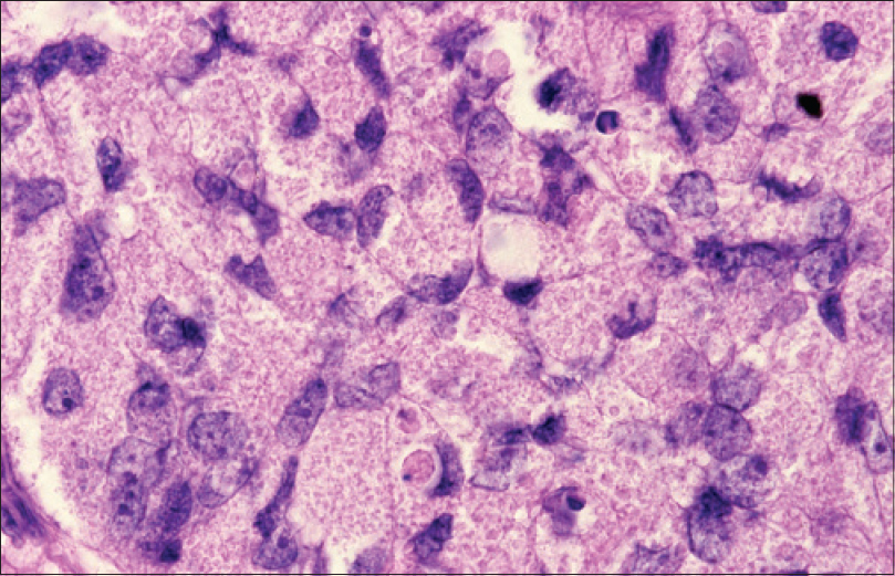
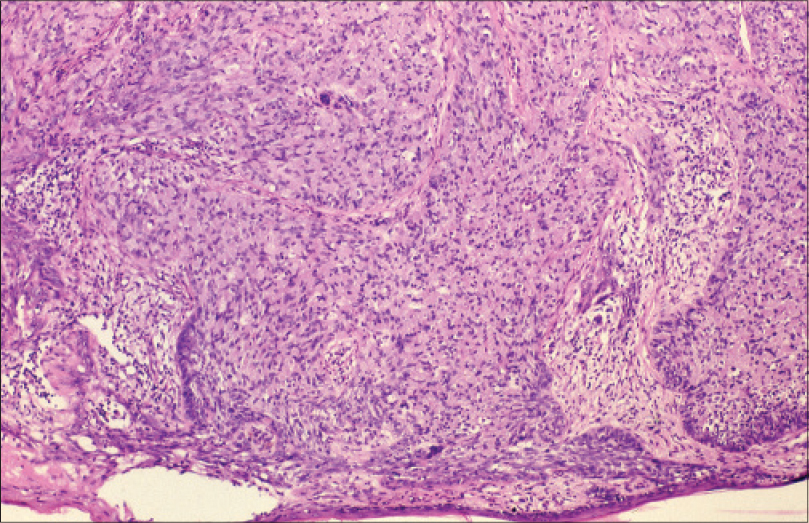
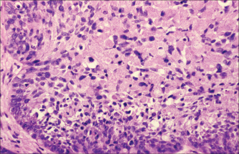
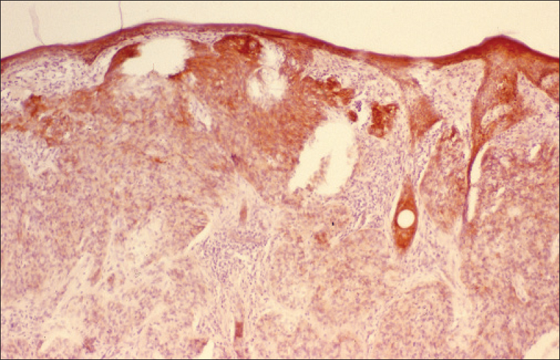
Microscopic findings showed a superficially ulcerated tumor containing solid deposits of polygonal cells with voluminous cytoplasm filled with eosinophilic granules [Figure - 2]. The periphery of the solid deposits was lined with palisades of low columnar basophilic cells [Figure - 3] and [Figure - 4]. Mitoses were present and dispersed. Solar keratosis was seen in proximity to the tumor. Immunohistochemistry of the tumor cells revealed cytokeratin expression [Figure - 5]. S-100 protein and neuron specific enolase (NSE) were negative. The patient was followed up for several years with no local recurrence of the tumor.
 |
| Figure 2: Granular cells (H and E, 400×) |
 |
| Figure 3: Solid granular cell structures with segments of cellular palisades on the periphery (H and E, 100×) |
 |
| Figure 4: Solid granular cell structures with segments of cellular palisades on the periphery (H and E, 200×) |
 |
| Figure 5: Cytokeratin expression AE1/AE3 (100×) |
Granular cell basal cell carcinoma is an extremely rare histological variant that was first described in 1979. [2] In all the reported cases to date, only a solitary lesion has been described with one exception where multiple lesions were seen. [13] Due to the small number of observed cases, epidemiologic and prognostic data evaluation is only tentative; however, Kanitakis and Chouvet attempted to do so in their study. [11] In the reported cases, including our case, the clinical picture, course and incidence of most granular cell basal cell carcinomas on the sun-exposed areas were no different from common basal cell carcinoma. The majority of granular cell basal cell carcinomas developed on the face and in the remaining cases on the chest.
Granular cell basal cell carcinoma does not require a different therapeutic approach than is used in usual variants of basal cell carcinoma. Nevertheless, the correct diagnosis is important because the granular cells may also be present in other tumors. The histological picture is characteristic with the formation of solid deposits of granular cells; the periphery is lined with cylindrical basophilic cells, as is typical for other variants of basal cell carcinoma. Positive expression of cytokeratin clearly confirms the epithelial origin. [1],[5],[11],[12]
Due to a partial convergence of some microscopic features, it is necessary to distinguish a granular cell tumor whose cells do not express cytokeratin but express S-100 protein and neuron specific enolase. [1] In broadening the differential diagnosis, one could also consider leiomyosarcoma and angiosarcoma, which are characterized by the expression of muscle and endothelial markers, respectively. Granular cell ameloblastoma with its typical localization does not represent a diagnostic problem. [1],[11]
Immunohistochemical studies performed to clarify the origin of cytoplasmic granules in granular cell basal cell carcinoma and in other granular cell tumors suggest their lysosomal genesis, probably due to degenerative cellular processes. [7],[11] Electron microscopic findings confirm this hypothesis.
| 1. |
Weedon D. Skin Pathology. Edinburgh: Churchill Livingstone; 2002.
[Google Scholar]
|
| 2. |
Bar RJ, Graham JH. Granular cell basal cell carcinoma. A distinct histopathologic entity. Arch Dermatol 1979;115:1064-7.
[Google Scholar]
|
| 3. |
Mrak RE, Baker GF. Granular cell basal cell carcinoma. J Cutan Pathol 1987;14:37-42.
[Google Scholar]
|
| 4. |
Le Boit PE, Barr RJ, Burall S, Metcalf JS, Yen TS, Wick MR. Primitive polypoid granular-cell tumor and other cutaneous granular-cell neoplasms of apparent nonneural origin. Am J Surg Pathol 1991;15:48-58.
[Google Scholar]
|
| 5. |
Garcia Prats MD, López Carrara M, Martinez-González MA, Ballestin C, Gil R, De Prada I. Granular cell basal cell carcinoma. Light microscopy, immunohistochemical and ultrastructural study. Virchows Arch A Pathol Anat Histopathol 1993;422:173-7.
[Google Scholar]
|
| 6. |
Reichel M. Granular cell basal cell carcinoma. Cutis 1997;59:88-90.
[Google Scholar]
|
| 7. |
Boscaino A, Tornillo L, Orabona P, Staibano S, Gentile R, De Rosa G. Granular cell basal cell carcinoma of the skin. Report of a case with immunohistochemical positivity for lysozyme. Tumori 1997;83:712-4.
[Google Scholar]
|
| 8. |
Hayden AA, Shamma HN. Ber-EP4 and MNF-116 in a previously undescribed morphologic pattern of granular basal cell carcinoma. Am J Dermatopathol 2001;23:530-2.
[Google Scholar]
|
| 9. |
Myung J, Ha S, Park C, Kang S, Kim S. A case of granular basal cell carcinoma. Korean J Dermatol 2002;40:1128-31.
[Google Scholar]
|
| 10. |
Dundr P, Stork J, Povýsil C, Vosmik F. Granular cell basal cell carcinoma. Australas J Dermatol 2004;45:70-2.
[Google Scholar]
|
| 11. |
Kanitakis J, Chouvet B. Granular-cell basal cell carcinoma of the skin. Eur J Dermatol 2005;15:301-3.
[Google Scholar]
|
| 12. |
Claassen SL, Royer MC, Rush WL. Granular cell basal cell carcinoma: Report of a Case and Review of the Literature. Am J Dermatopathol 2014;36:e121-4.
[Google Scholar]
|
| 13. |
Jedrych J, Busam KJ. Multiple lesions of granular cell basal cell carcinoma: A case report. J Cutan Pathol 2014;41:45-50.
[Google Scholar]
|
Fulltext Views
3,531
PDF downloads
2,490


