Translate this page into:
Hemorrhagic herpes zoster: A rare presentation
Corresponding author: Dr. Sonal Fernandes, Department of Dermatology, Venereology and Leprosy, Father Muller Medical College, Mangalore - 575 002, Karnataka, India. sonferns007@gmail.com
-
Received: ,
Accepted: ,
How to cite this article: Fernandes S, D’Souza M, Nandakishore B. Hemorrhagic herpes zoster: A rare presentation. Indian J Dermatol Venereol Leprol 2023;89:292-3.
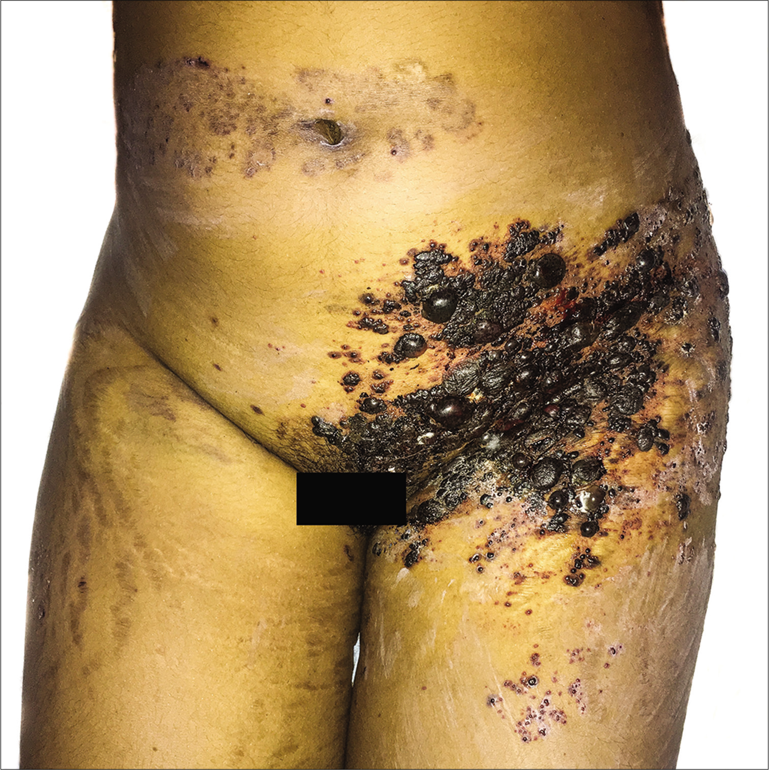
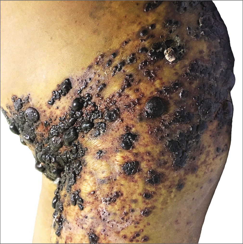
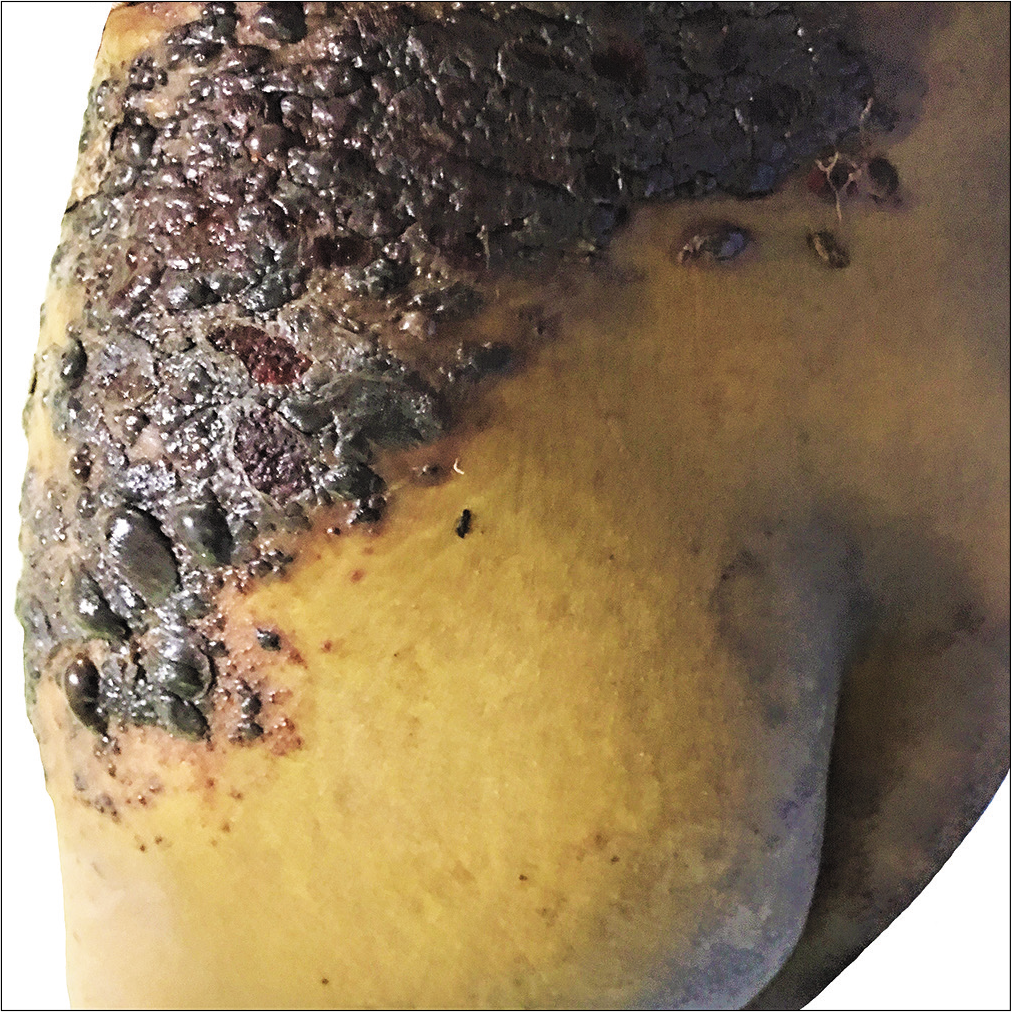
A 20-year-old woman presented with a 2-day history of multiple, painful, fluid-filled lesions resembling a bunch of grapes over her lower abdomen and back. She is a known case of autoimmune hemolytic anemia - Evans syndrome - receiving oral prednisolone for last 1 year. Cutaneous examination revealed closely grouped hemorrhagic bullae and vesicles over her left twelfth thoracic and first two lumbar dermatomal segments [Figures 1a-c]. Blood biochemistry revealed platelet count- 2000/mm3, prothrombin time - 11 s, prothrombin time-international normalized ratio- 1.06 and activated partial thromboplastin time- 29 s. We made a diagnosis of hemorrhagic herpes zoster based on the characteristic clinical presentation. Immunosuppression and severe thrombocytopenia possibly led to hemorrhagic lesions. Treatment was initiated with oral valacyclovir, oral and topical antibiotics and regular dressings. Supportive therapy with oral steroids and platelet transfusions were administered for treating the underlying autoimmune hemolytic anemia. Following 1 week of antiviral therapy, the lesions started healing with erosions.

- Multiple hemorrhagic bullae and vesicles over the left abdomen and pelvic region

- Numerous grouped hemorrhagic vesicles and bullae noted over the left hip and upper thigh

- Lesions extending posteriorly to left lower back and gluteal region
Declaration of patient consent
The authors certify that they have obtained all appropriate patient consent.
Conflicts of interest
There are no conflicts of interest.
Financial support and sponsorship
Nil.


