Translate this page into:
High dynamic range conversion and contrast adjustment to improve quality of dermoscopy imaging in vitiligo: A pilot study
2 Department of Dermatology, KIMS Al Shifa Hospital, Perinthalmana, Malappuram, Kerala, India
Correspondence Address:
Feroze Kaliyadan
Department of Dermatology, College of Medicine, King Faisal University, Al-Ahsa 31982
Saudi Arabia
| How to cite this article: Kaliyadan F, Ashique KT. High dynamic range conversion and contrast adjustment to improve quality of dermoscopy imaging in vitiligo: A pilot study. Indian J Dermatol Venereol Leprol 2019;85:682 |
Sir,
Dermoscopy is being increasingly used as a tool to assess the stages of evolution and the disease activity in vitiligo.[1],[2] Conversion of normal dermoscopy images to high dynamic range images has been performed to improve the quality of imaging in the context of pigmented skin tumors.[3],[4] We aimed to evaluate the effectiveness of high dynamic range conversion in improving visualization of dermoscopy features in vitiligo.
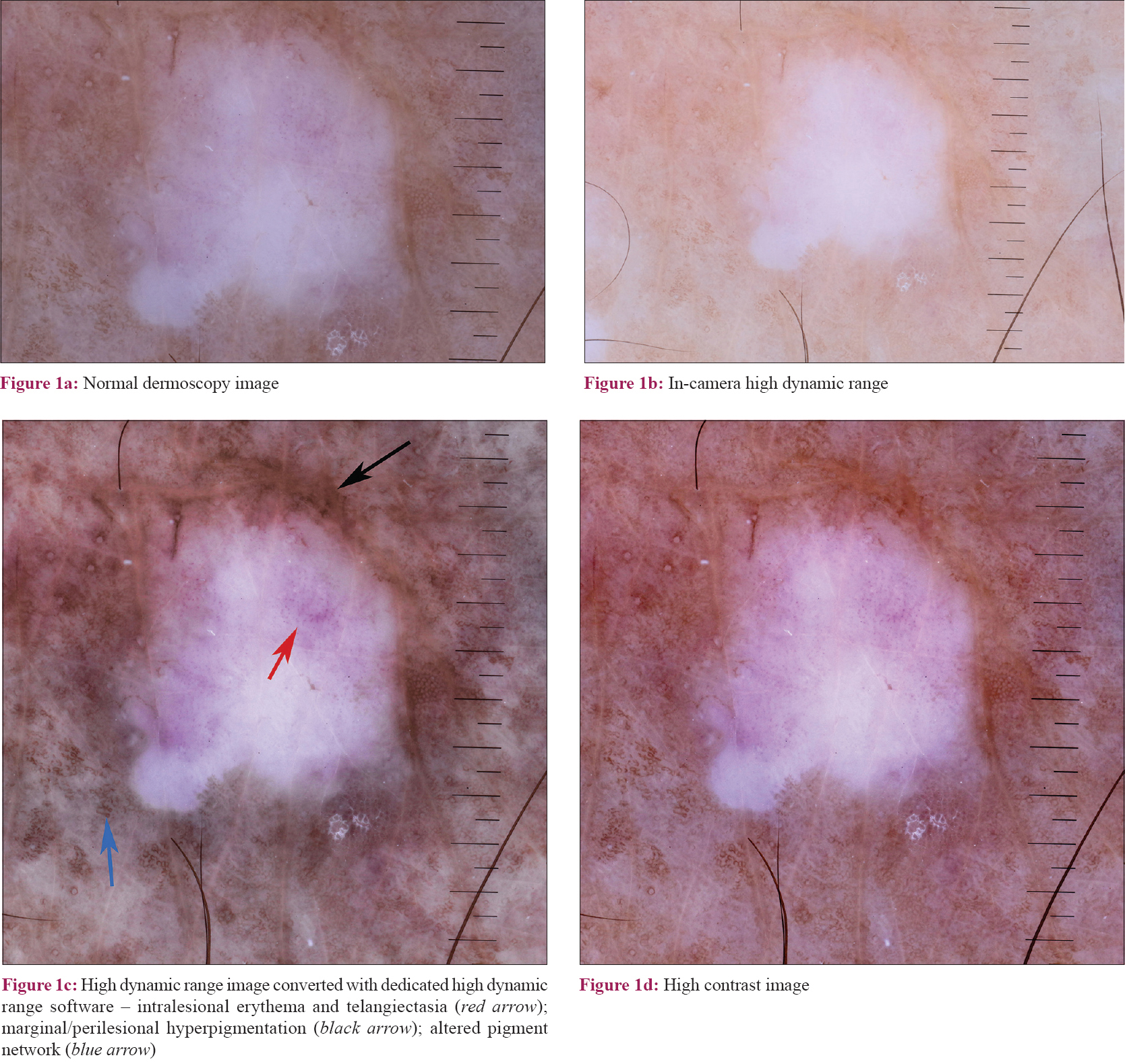
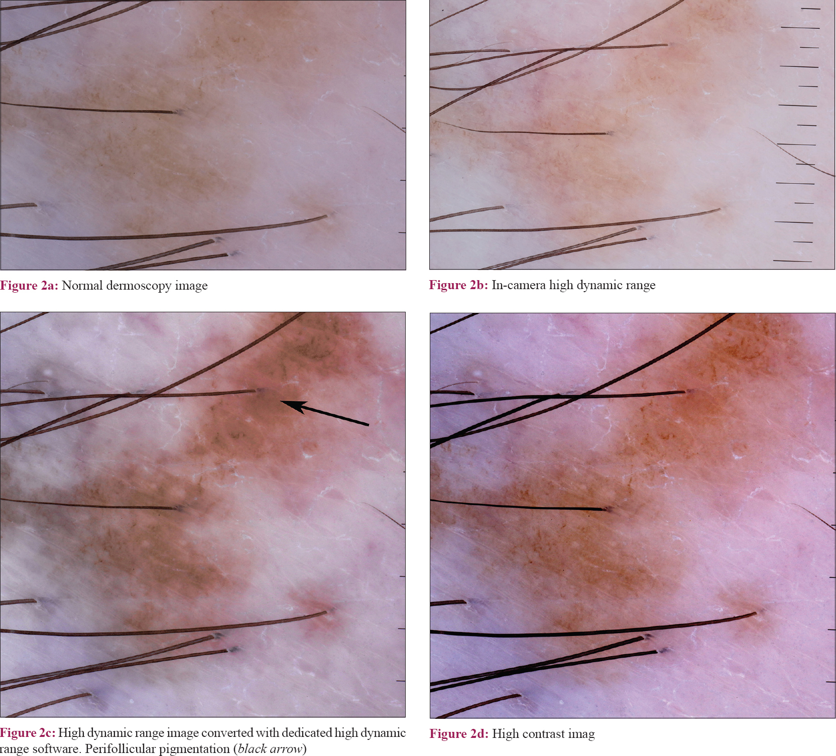
In this study, 20 vitiligo lesions in six patients (all cases of unstable vitiligo) were studied. Dermoscopy images (polarized 10×) were captured using a Dermlite® Foto ii Pro dermoscope attached to a Canon® 650D digital SLR camera. Two high dynamic range images were obtained – one from the in-built high dynamic range conversion system on the camera and another one using Topaz® software that converts the original dermoscopy image to high dynamic range. A high-contrast image was obtained by increasing the contrast parameter of the original dermoscopy image. All editing works were done using Adobe Photoshop CS6® (high dynamic range software was used as a plugin). Conversion parameters were kept uniform for all sets of images (for high dynamic range, dynamic pop II was used in Topaz® and for contrast enhancement, a fixed 50% increase was used) [Figure - 1] and [Figure - 2].
 |
| Figure 1: |
 |
| Figure 2: |
Dermoscopy images were viewed by two dermatologists who were blinded to the label, and for each image the quality of visualization of dermoscopy features was graded on a global scale of 1–5 (1 = lowest quality to 5 = highest quality). For statistical analysis, Kruskal–Wallis test was used to compare the mean ratings across the four groups.
The highest mean ranks were obtained for the images converted using dedicated high dynamic range software (mean rank = 61.3), followed by the high-contrast images (mean rank = 59.3). The lowest mean ranks were obtained for the "in-camera" high dynamic range conversion (mean rank = 12), and the mean ranks for the raw dermoscopy image was 29.4. These results were statistically significant (Kruskal–Wallis, degrees of freedom = 3, H = 63.7, P value < 0.001).
The dynamic range of a digital image refers to the ratio between the maximum and minimum detectable light intensities.[3] High dynamic range conversion involves increasing this range of luminosity. Classically, high dynamic range images are made by combining images of at least three different exposures (one normal exposure, one under and one over-exposed image).[3] However, there are softwares that can convert single image into high dynamic range, although the results are in general not as good as combinations of images with true differential exposures. The dermoscopic features of vitiligo mainly involve changes in the pigmentary pattern, particularly in the perifollicular area.[1],[2] The conversion of dermoscopy images to high dynamic range can help visualize these patterns better. The use of dedicated high dynamic range software might be limited by the cost and unfamiliarity of dermatologists with the concept. Our study suggests that even a simple contrast enhancement of the images can improve visualization of dermoscopic features in vitiligo and this can be easily done with any basic photo-editing software. In-camera, high dynamic range quality might be variable across cameras and further studies are warranted to study this aspect. It is possible that the high dynamic range algorithm in the camera we used is more suitable for specific situations such as landscape or architectural photography and less effective for dermoscopic imaging. The small sample size is the major limitation of the study. The global rating and only two evaluators being involved were the other limitations.
To conclude, high dynamic range conversion of dermoscopy images is a technique that can be effective in improving quality of visualization of dermoscopic features in vitiligo. Interestingly, for vitiligo, even simple contrast enhancement can produce significant improvement in the quality of dermoscopy imaging.
Financial support and sponsorship
Nil.
Conflicts of interest
There are no conflicts of interest.
| 1. |
Kumar Jha A, Sonthalia S, Lallas A, Chaudhary RK. Dermoscopy in vitiligo: Diagnosis and beyond. Int J Dermatol 2018;57:50-4.
[Google Scholar]
|
| 2. |
Thatte SS, Khopkar US. The utility of dermoscopy in the diagnosis of evolving lesions of vitiligo. Indian J Dermatol Venereol Leprol 2014;80:505-8.
[Google Scholar]
|
| 3. |
Braun RP, Marghoob A. High-dynamic-range dermoscopy imaging and diagnosis of hypopigmented skin cancers. JAMA Dermatol 2015;151:456-7.
[Google Scholar]
|
| 4. |
Sato T, Tanaka M. A case of a superficial spreading melanoma in situ diagnosed via digital dermoscopic monitoring with high dynamic range conversion. Dermatol Pract Concept 2014;4:57-60.
[Google Scholar]
|
Fulltext Views
2,136
PDF downloads
2,195


