Generic selectors
Exact matches only
Search in title
Search in content
Post Type Selectors
Search in posts
Search in pages
Filter by Categories
15th National Conference of the IAOMFP, Chennai, 2006
Abstract
Abstracts from current literature
Acne in India: Guidelines for management - IAA Consensus Document
Addendum
Announcement
Art & Psychiatry
Article
Articles
Association Activities
Association Notes
Award Article
Book Review
Brief Report
Case Analysis
Case Letter
Case Letters
Case Notes
Case Report
Case Reports
Clinical and Laboratory Investigations
Clinical Article
Clinical Studies
Clinical Study
Commentary
Conference Oration
Conference Summary
Continuing Medical Education
Correspondence
Corrigendum
Cosmetic Dermatology
Cosmetology
Current Best Evidence
Current Issue
Current View
Derma Quest
Dermato Surgery
Dermatopathology
Dermatosurgery Specials
Dispensing Pearl
Do you know?
Drug Dialogues
e-IJDVL
Editor Speaks
Editorial
Editorial Remarks
Editorial Report
Editorial Report - 2007
Editorial report for 2004-2005
Errata
Erratum
Focus
Fourth All India Conference Programme
From Our Book Shelf
From the Desk of Chief Editor
General
Get Set for Net
Get set for the net
Guest Article
Guest Editorial
History
How I Manage?
IADVL Announcement
IADVL Announcements
IJDVL Awards
IJDVL AWARDS 2015
IJDVL Awards 2018
IJDVL Awards 2019
IJDVL Awards 2020
IJDVL International Awards 2018
Images in Clinical Practice
Images in Dermatology
In Memorium
Inaugural Address
Index
Knowledge From World Contemporaries
Leprosy Section
Letter in Response to Previous Publication
Letter to Editor
Letter to the Editor
Letter to the Editor - Case Letter
Letter to the Editor - Letter in Response to Published Article
LETTER TO THE EDITOR - LETTERS IN RESPONSE TO PUBLISHED ARTICLES
Letter to the Editor - Observation Letter
Letter to the Editor - Study Letter
Letter to the Editor - Therapy Letter
Letter to the Editor: Articles in Response to Previously Published Articles
Letters in Response to Previous Publication
Letters to the Editor
Letters to the Editor - Letter in Response to Previously Published Articles
Letters to the Editor: Case Letters
Letters to the Editor: Letters in Response to Previously Published Articles
Media and news
Medicolegal Window
Messages
Miscellaneous Letter
Musings
Net Case
Net case report
Net Image
Net Images
Net Letter
Net Quiz
Net Study
New Preparations
News
News & Views
Obituary
Observation Letter
Observation Letters
Oration
Original Article
ORIGINAL CONTRIBUTION
Original Contributions
Pattern of Skin Diseases
Pearls
Pediatric Dermatology
Pediatric Rounds
Perspective
Presedential Address
Presidential Address
Presidents Remarks
Quiz
Recommendations
Regret
Report
Report of chief editor
Report of Hon : Treasurer IADVL
Report of Hon. General Secretary IADVL
Research Methdology
Research Methodology
Resident page
Resident's Page
Resident’s Page
Residents' Corner
Residents' Corner
Residents' Page
Retraction
Review
Review Article
Review Articles
Reviewers 2022
Revision Corner
Self Assessment Programme
SEMINAR
Seminar: Chronic Arsenicosis in India
Seminar: HIV Infection
Short Communication
Short Communications
Short Report
Snippets
Special Article
Specialty Interface
Studies
Study Letter
Study Letters
Supplement-Photoprotection
Supplement-Psoriasis
Symposium - Contact Dermatitis
Symposium - Lasers
Symposium - Pediatric Dermatoses
Symposium - Psoriasis
Symposium - Vesicobullous Disorders
SYMPOSIUM - VITILIGO
Symposium Aesthetic Surgery
Symposium Dermatopathology
Symposium-Hair Disorders
Symposium-Nails Part I
Symposium-Nails-Part II
Systematic Review and Meta-Analysis
Systematic Reviews and Meta-analyses
Systematic Reviews and Meta-analysis
Tables
Technology
Therapeutic Guideline-IADVL
Therapeutic Guidelines
Therapeutic Guidelines - IADVL
Therapeutics
Therapy
Therapy Letter
Therapy Letters
View Point
Viewpoint
What’s new in Dermatology
View/Download PDF

Translate this page into:

Observation Letter
88 (
3
); 409-412
doi:
10.25259/IJDVL_688_2021
pmid:
35434989

Iatrogenic Kaposi’s sarcoma induced by rituximab and corticosteroid treatment for pemphigus vegetans in an HIV-negative patient

Department of Dermatology, Mohammed VI University Hospital, Oujda, Morocco
Laboratory of Epidemiology, Clinical Research and Public Health, Medical School of Oujda, Mohammed First University, Oujda, Morocco

Corresponding author: Dr. Hanane Daflaoui, Department of Dermatology, Mohammed VI University Hospital, Mohammed First University, Oujda, Morocco. hanane.daflaoui@gmail.com

Licence
This is an open-access article distributed under the terms of the Creative Commons Attribution-Non Commercial-Share Alike 4.0 License, which allows others to remix, transform, and build upon the work non-commercially, as long as the author is credited and the new creations are licensed under the identical terms.

How to cite this article: Daflaoui H, Saddouk H, Ouadi I, Zizi N, Dikhaye S. Iatrogenic Kaposi’s sarcoma induced by rituximab and corticosteroid treatment for pemphigus vegetans in an HIV-negative patient. Indian J Dermatol Venereol Leprol 2022;88:409-12.

Sir

Kaposi’s sarcoma is a rare multifocal angioproliferative tumour caused by human herpesvirus-8 infection. Four clinical variants have been distinguished: classic Kaposi’s sarcoma, endemic Kaposi’s sarcoma, AIDS-related Kaposi’s sarcoma and iatrogenic Kaposi’s sarcoma (an entity that usually affects patients who undergo long-term immunosuppressant therapy for immune-mediated or proliferative disorders or solid-organ transplantations).1

An eighty two-year-old Moroccan man with a history of diabetes mellitus, hypertension and treated pulmonary tuberculosis attended the dermatology department of Mohammed VI University Hospital of Oujda with multiple purpuric lesions over the legs since three months. He was on oral prednisone 55 mg/day for histopathologically proven pemphigus vegetans. He was also receiving gliclazide 30 mg/day and amlodipine 5 mg/day for diabetes mellitus and hypertension, respectively. Due to inadequate response of pemphigus vegetans to treatment with systemic corticosteroids, he was administered two doses of 1g rituximab 15 days apart, which allowed slow tapering of oral prednisone. Six months after the initiation of corticosteroids and nearly three months after the first dose of rituximab, the patient noticed the appearance of multiple brightly erythematous papules as well as purpuric lesions on the lower limbs.

Clinical examination revealed multiple purple and dusky erythematous papules, plaques and nodules on the anterior aspect of the right leg [Figure 1a] and around the left ankle. Most of the lesions were scaly while some lesions on the left ankle were ulcerated [Figure 1b]. Similar purple papules were also seen on the right thigh [Figure 1c] hips and trunk along with purpuric lesions on the thighs. His pemphigus was in remission. Dermoscopy of the papules and plaques showed purple-pink peripheral scales, small brown globules and a rainbow pattern [Figures 2a-2c].
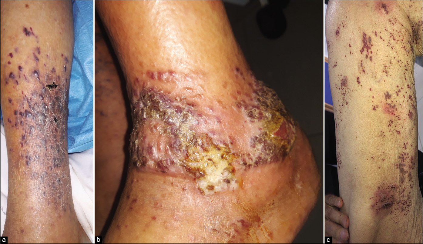
(a) Purple and dusky erythematous papules, plaques and nodules of Kaposi’s sarcoma on the right leg, (b) Kaposi’s sarcoma manifesting as purple and erythematous papules and plaques with scaling and ulceration, (c) Purple papules of Kaposi’s sarcoma on the right thigh
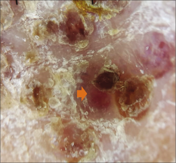
Figure 1:
(a) Purple and dusky erythematous papules, plaques and nodules of Kaposi’s sarcoma on the right leg, (b) Kaposi’s sarcoma manifesting as purple and erythematous papules and plaques with scaling and ulceration, (c) Purple papules of Kaposi’s sarcoma on the right thigh
Dermoscopy of a nodular lesion of Kaposi’s sarcoma showing nodular lesion with peripheral scales {orange arrow} (3Gen DermLite III DL3N, polarised light, ×10)
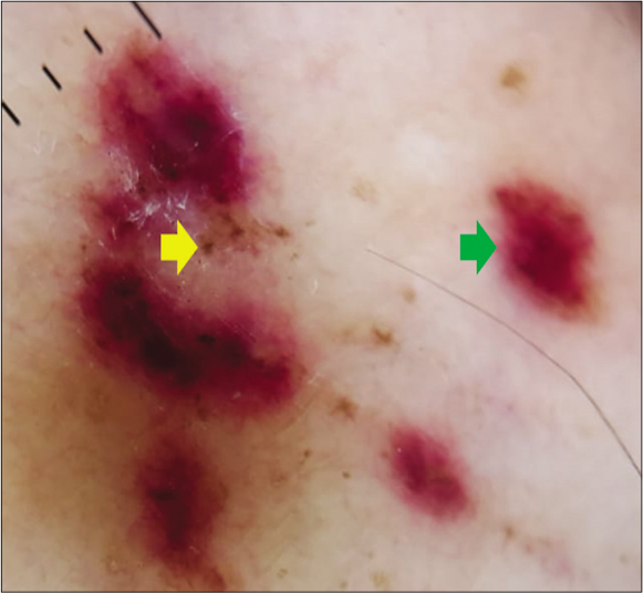
Figure 2a:
Dermoscopy of a nodular lesion of Kaposi’s sarcoma showing nodular lesion with peripheral scales {orange arrow} (3Gen DermLite III DL3N, polarised light, ×10)
Dermoscopy of a nodular lesion of Kaposi’s sarcoma showing small brown globules {yellow arrow} and purple-pink colouration {green arrow} (3Gen DermLite III DL3N, polarised light, ×10)
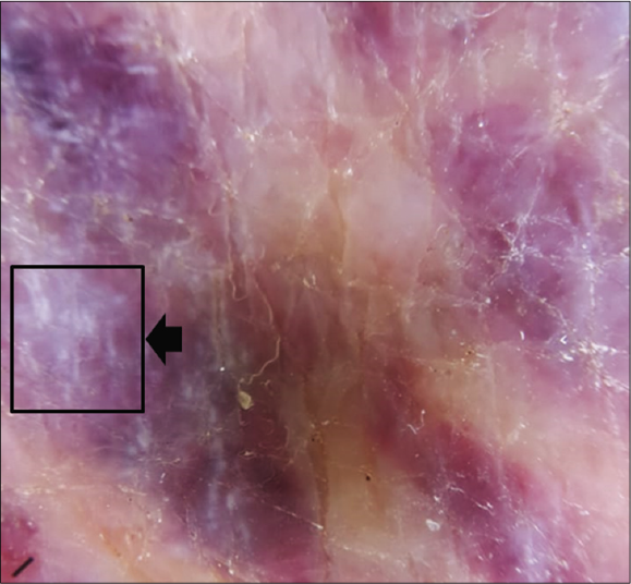
Figure 2b:
Dermoscopy of a nodular lesion of Kaposi’s sarcoma showing small brown globules {yellow arrow} and purple-pink colouration {green arrow} (3Gen DermLite III DL3N, polarised light, ×10)
Dermoscopy of a nodular lesion of Kaposi’s sarcoma showing rainbow pattern {black arrow} (3Gen DermLite III DL3N, polarised light, ×10)
Figure 2c:
Dermoscopy of a nodular lesion of Kaposi’s sarcoma showing rainbow pattern {black arrow} (3Gen DermLite III DL3N, polarised light, ×10)

Skin biopsy from a purple nodule on the right leg showed circumscribed proliferation of spindle cells arranged in fascicles, slit-like vascular channels [Figure 3a], extravasation of erythrocytes [Figure 3b] and lymphocytic inflammatory infiltrate. The nuclei of the tumour cells demonstrated immunoreactivity for CD31 [Figure 4a] and latent nuclear antigen-1 of human herpesvirus-8 [Figure 4b]. Indirect immunofluorescence detected antibodies to human herpesvirus-8 lytic antigens. Serology for HIV was negative.
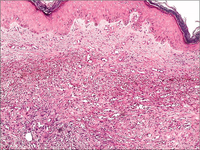
Skin biopsy from a nodule of Kaposi’s sarcoma on the right leg showing circumscribed proliferation of spindle cells arranged in fascicles and slit-like vascular channels (H and E, ×50)
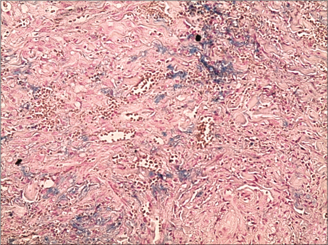
Figure 3a:
Skin biopsy from a nodule of Kaposi’s sarcoma on the right leg showing circumscribed proliferation of spindle cells arranged in fascicles and slit-like vascular channels (H and E, ×50)
Skin biopsy from a nodule of Kaposi’s sarcoma on the right leg showing circumscribed proliferation of spindle cells arranged in fascicles, slit-like vascular channels and extravasation of erythrocytes (Perl’s stain, ×200)
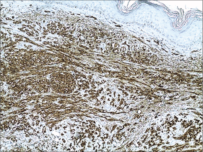
Figure 3b:
Skin biopsy from a nodule of Kaposi’s sarcoma on the right leg showing circumscribed proliferation of spindle cells arranged in fascicles, slit-like vascular channels and extravasation of erythrocytes (Perl’s stain, ×200)
The nuclei of the tumour cells of Kaposi’s sarcoma demonstrating immunoreactivity for CD31 (13B10 clone ×200)
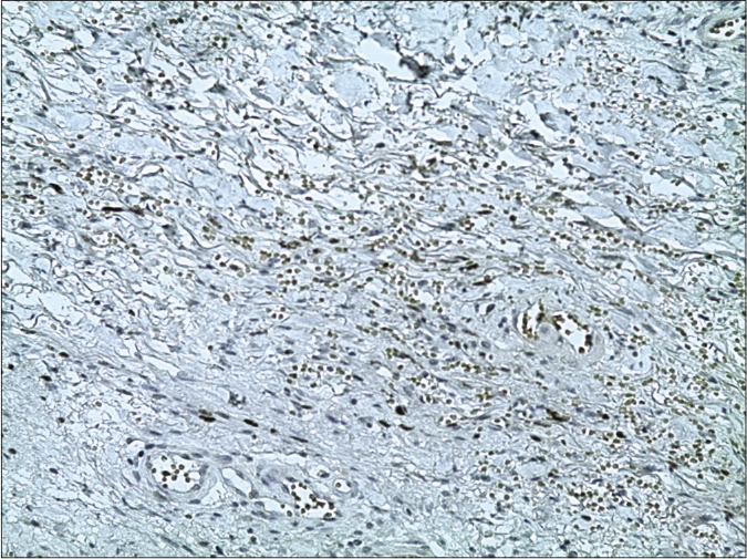
Figure 4a:
The nuclei of the tumour cells of Kaposi’s sarcoma demonstrating immunoreactivity for CD31 (13B10 clone ×200)
The nuclei of the tumour cells of Kaposi’s sarcoma demonstrating immunoreactivity for latent nuclear antigen-1 of human herpesvirus-8 (13B10 clone, latent nuclear antigen-1 of human herpesvirus-8, ×200)
Figure 4b:
The nuclei of the tumour cells of Kaposi’s sarcoma demonstrating immunoreactivity for latent nuclear antigen-1 of human herpesvirus-8 (13B10 clone, latent nuclear antigen-1 of human herpesvirus-8, ×200)

A whole body computed tomography scan and upper gastrointestinal endoscopy and colonoscopy showed no evidence of metastasis. A final diagnosis of nodular iatrogenic Kaposi’s sarcoma was made and the patient was treated with topical 5-fluorouracil. Systemic corticosteroids were continued to be tapered as per the clinical response. Marked resolution of skin lesions was noted after three months of once-daily application of 5-fluorouracil [Figure 5a-c].
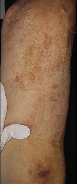
Clinical images after treatment: Regression of Kaposi lesions of the right thigh
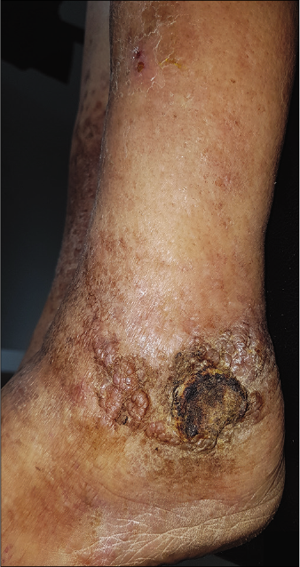
Figure 5a:
Clinical images after treatment: Regression of Kaposi lesions of the right thigh
Clinical images after treatment: Partial regression of Kaposi lesions of the left ankle
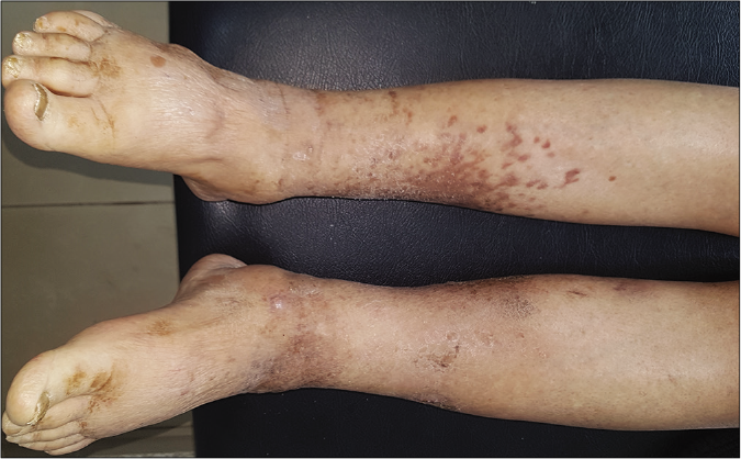
Figure 5b:
Clinical images after treatment: Partial regression of Kaposi lesions of the left ankle
Clinical images after treatment: Partial regression of Kaposi lesions of the right leg
Figure 5c:
Clinical images after treatment: Partial regression of Kaposi lesions of the right leg

Kaposi’s sarcoma typically presents as purple patches and plaques with a possible involvement of extracutaneous sites, especially the oral mucosa, gastrointestinal tract and lymph nodes. Since its first description, four clinical variants of Kaposi’s sarcoma have been described: classic, endemic, iatrogenic and AIDS related.1,2 The iatrogenic variant of Kaposi’s sarcoma is rarely reported and accounts for 5% to 20% of cases in the previous reported series.3-6 It is most often seen in patients having undergone solid organ transplantation and those undergoing prolonged immunosuppressive therapy with agents such as cyclosporine, azathioprine, corticosteroids and rituximab.3

Our patient was dually immunosuppressed with corticosteroids and rituximab therapy. Rituximab is a chimeric monoclonal antibody which targets CD20 antigen expressed in more than 95% of normal and malignant B cells, inducing complement-mediated and antibody-dependent cellular cytotoxicity.

Several case reports and series have implicated rituximab in the development of Kaposi’s sarcoma in HIV-positive as well as HIV-negative patients. According to the recent studies, rituximab can decrease T-cell immunity;7,9 however, the link between rituximab-mediated CD20+ B-cell depletion and human herpesvirus-8 activation and the development of Kaposi’s sarcoma remain unclear. In all the previous reported cases, rituximab was administered along with other immunosuppressive agents which makes it difficult to delineate its exact role in the development of Kaposi’s sarcoma.7,8 Corticosteroids have also been implicated in the development and the dissemination of Kaposi’s sarcoma.10 Consequently, the role of corticosteroids in the causation of Kaposi’s sarcoma in our case cannot be excluded. Their mechanism of action is the inhibition of growth factor-beta, a protein that inhibits the growth of endothelial cells.10

There are no guidelines for the management of iatrogenic Kaposi’s sarcoma. Regardless, the reduction or the interruption of immunosuppressant agents is the most widely accepted therapeutic option often combined with local or systemic therapies depending on the disease stage and severity.11 In our case, given the stability of his pemphigus and the absence of new bullous lesions, it was decided to decrease the corticosteroid dose and not readminister rituximab. We opted for treatment with topical 5-fluorouracil for its proven anti-Kaposi’s sarcoma-associated herpes virus activity.12,13

This case report aims to increase awareness regarding the rare complication of Kaposi’s sarcoma following immunosuppressive treatment.

Declaration of patient consent

The authors certify that they have obtained all appropriate patient consent.

Financial support and sponsorship

Nil.

Conflicts of interest

There are no conflicts of interest.

References

  1. , , , , , . Kaposi sarcoma. Nat Rev Dis Primers. 2019;5:1-21.
    [CrossRef] [PubMed] [Google Scholar]
  2. , . Kaposi sarcoma updates. Dermatol Clin. 2019;4:505-17.
    [CrossRef] [PubMed] [Google Scholar]
  3. , . Epidemiology of Kaposi sarcoma : Review and description of the nonepidemic variant. Int J Dermatol. 2019;58:538-42.
    [CrossRef] [PubMed] [Google Scholar]
  4. , , , , . Demographic and histopathologic study of Kaposi's sarcoma in a dermatology clinic in the years of 2006 to 2011. Acta Med Iran. 2014;52:381-4.
    [Google Scholar]
  5. , , , , , , et al. Kaposi's sarcoma : A population-based cancer registry descriptive study of 57 consecutive cases diagnosed between 1977 and 2009. Int J Dermatol. 2014;53:549.e-54.
    [CrossRef] [PubMed] [Google Scholar]
  6. , , , , , , et al. Factors affecting progression-free survival in non-HIV-related Kaposi sarcoma. J Dermatol Treat. 2016;27:275-7.
    [CrossRef] [PubMed] [Google Scholar]
  7. , , , . Kaposi's sarcoma after autologous stem-cell transplantation and rituximab treatment. J Oncol Pract. 2018;14:565.
    [CrossRef] [PubMed] [Google Scholar]
  8. , , . Kaposi sarcoma associated with rituximab-based cytotoxic therapy. J Oncol Pharm Pract. 2020;26:220-3.
    [CrossRef] [PubMed] [Google Scholar]
  9. , , , , , , et al. Kaposi's sarcoma : Aetiopathogenesis, histology and clinical features. JEADV. 2003;17:138-54.
    [CrossRef] [PubMed] [Google Scholar]
  10. , , , , , , et al. Glucocorticoids induce Kaposi's sarcoma cell proliferation through the regulation of transforming growth factor-β. Blood Am J Hematol. 1997;89:1491-500.
    [CrossRef] [Google Scholar]
  11. , , . Iatrogenic Kaposi's sarcoma : A retrospective cohort study in an Italian tertiary care centre. Clin Oncol. 2017;29:e165-71.
    [CrossRef] [PubMed] [Google Scholar]
  12. , , . Pemetrexed inhibits Kaposi's sarcoma-associated herpesvirus replication through blocking dTMP synthesis. Antivir Res. 2020;180:104825.
    [CrossRef] [PubMed] [Google Scholar]
  13. , , , , . Kaposi's sarcoma resolves after sirolimus therapy in a patient with pemphigus vulgaris. Arch Dermatol. 2008;144:654-7.
    [CrossRef] [PubMed] [Google Scholar]

Fulltext Views
4,738

PDF downloads
3,873
View/Download PDF
Download Citations
BibTeX
RIS
Show Sections