Translate this page into:
Late-onset apocrine chromhidrosis
2 Departments of Dermatology, Izmir Katip �elebi University, School of Medicine, Izmir, Turkey
3 Department of Pathology, Izmir Atat�rk Research and Traning Hospital, Izmir, Turkey
Correspondence Address:
Ilgul Bilgin
149, Sok. 1/16 Karaba?lar, Hatay, Izmir
Turkey
| How to cite this article: Bilgin I, Kelekci KH, Catal S, Calli A. Late-onset apocrine chromhidrosis . Indian J Dermatol Venereol Leprol 2014;80:579 |
Sir,
Apocrine chromhidrosis is a rare condition characterized by the secretion of colored sweat. Apocrine sweat glands are located in the axillae, anogenital skin, areolae and over the skin of the trunk, face and scalp. It has been demonstrated that lipofuscin pigment, produced by apocrine glands is responsible for the colored sweat. The color of apocrine sweat may be brown, yellow, green, blue or black depending on the varying concentrations or oxidation states of the lipofuscin granules. Darker colors are due to higher states of oxidation. There is no gender or geographic predisposition and chromhidrosis is not influenced by seasonal factors. [1] We describe an elderly male with acquired chromhidrosis.
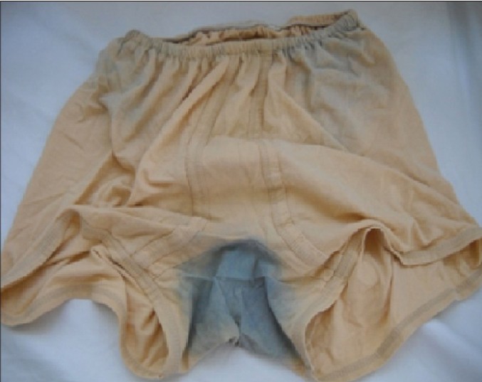
A 76-year-old man presented with a 4-month history of blue staining of clothing over his groins, back and axillae. He complained of blue sweat that was evident after physical activity. General and systemic examinations were normal. On examination, there was an odorless blue discoloration on his inner garments [Figure - 1]. On dermatological examination, skin appeared normal. Routine laboratory investigations were normal. Culture of skin scrapings revealed normal skin flora. Histopathological examination of the skin biopsy showed dilated apocrine acini with bluish secretion [Figure - 2], and periodic acid-Schiff (PAS) positive granules in the cytoplasm of acinar apocrine cells [Figure - 3]. Based on the history, clinical evaluation and laboratory workup, a diagnosis of apocrine chromhidrosis was made. The patient was offered botulinum toxin and capsaicin treatment but he did not accept it.
 |
| Figure 1: Blue-stained garment |
 |
| Figure 2: Dilated apocrine acini with bluish secretion (H and E×200) |
 |
| Figure 3: PAS positive granules in the cytoplasm of acinar apocrine cells (PAS stain ×400) |
Apocrine chromhidrosis may sometimes be difficult to differentiate from eccrine chromhidrosis and pseudochromhidrosis. Eccrine chromhidrosis occurs due to water soluble pigment excreted from eccrine glands which may occur after ingestion of certain dyes or drugs. Pseudochromhidrosis occurs when colorless eccrine sweat becomes colored on the surface of the skin as a result of extrinsic dyed clothing, paints, or chromogenic bacteria. Fungal and bacteriologic cultures are necessary to exclude infectious causes of pseudochromhidrosis. The presence of lipofuscin granules in apocrine cells on histological examination usually confirms the diagnosis of apocrine chromhidrosis. [2]
Although the disorder is benign in nature, the appearance of colored sweat may oblige patients to change their clothing multiple times during the day as a result of the discoloration that ensues. The production of colored sweat frequently has an impact on psychological and social functioning. [3],[4] Apocrine sweat glands respond to emotional stimuli such as anxiety, pain or sexual arousal. Our patient gave a history of intense emotional stress which might be a possible reason for his disease.
Treatment options for this entity are limited but temporary relief with capsaicin cream and botulinum toxin has been reported. However, clinical relapse occurs when therapy is stopped. It is important to reassure patients that apocrine chromhidrosis is a benign condition. [4]
The prevalence and average age of onset of apocrine chromhidrosis is not well documented. It may develop at any age but is more often noticed after puberty, when apocrine secretory function is activated. Male to female ratio is unknown because there are too few cases reported to draw any meaningful conclusions. [3] The age of the patients reported in the literature range from 9 months [5] to 62 years. [3] A 60-year-old postmenopausal female with bluish secretion was reported by Mapare et al. [2] One of the oldest patients with chromhidrosis was a 62-year-old woman, with a complaint of ebony-colored sweat. [3] We were unable to find reports of any patient older than our 76-year-old patient.
| 1. |
Griffith JR. Isolated areolar apocrine chromhidrosis. Pediatrics 2005;115:e239-41.
[Google Scholar]
|
| 2. |
Mapare A, Tapre VN, Khandelwal AK. Apocrine chromhidrosis over dorsum of foot: A case report. IOSR J Dent Med Sci 2012;2:33-4.
[Google Scholar]
|
| 3. |
Beer K, Oakley H. Axillary chromhidrosis: Report of a case, review of the literature and treatment considerations. Cosmet Dermatol 2010;9:318-20.
[Google Scholar]
|
| 4. |
Polat M, Dikilitas M, Gözübüyükoðullarý A, Alli N. Apocrine chromhidrosis. Clin Exp Dermatol 2009;34:373-4.
[Google Scholar]
|
| 5. |
Carman KB, Aydogdu SD, Sabuncu I, Yarar C, Yakut A, Oztelcan B. Infant with chromhidrosis. Pediatr Int 2011;53:283-4.
[Google Scholar]
|
Fulltext Views
6,119
PDF downloads
2,316


