Translate this page into:
Linear and whorled nevoid hypermelanosis: A case report with dermoscopic findings
2 Department of Medical and Biological Sciences, Institution of Anatomic Pathology, Santa Maria della Misericordia University Hospital, Udine, Italy
Correspondence Address:
Giuseppe Stinco
Institute of Dermatology, Santa Maria della Misericordia University Hospital, Piazzale Santa Maria della Misericordia, 15-33100 Udine
Italy
| How to cite this article: Errichetti E, Pegolo E, Stinco G. Linear and whorled nevoid hypermelanosis: A case report with dermoscopic findings. Indian J Dermatol Venereol Leprol 2016;82:91-93 |
Sir,
Linear and whorled nevoid hypermelanosis is a rare pigmentary disorder presenting with brownish macules in a streaky configuration along the lines of Blaschko; the macules lack atrophy and are not preceded by any inflammatory or palpable lesions.[1] The most common sites involved are the trunk and extremities; the palms, soles, face and mucosae are usually spared.[1],[2],[3] It is histopathologically characterized by an increase in basal layer pigmentation without melanocyte proliferation; pigment incontinence is generally absent.[1],[4] The lesions may be present at birth or may develop in the first few weeks of life and continue to progress for a year or two before stabilization.[2] Hyperpigmentation tends to persists throughout life, although the macules may become less prominent with age in some patients.[1],[2] Linear and whorled nevoid hypermelanosis does not show a sex predilection and typically occurs sporadically but rare familial instances have also been described.[1] It is considered to result from the expression of genetic mosaicism or chimerism and several cytogenetic alterations have been reported.[3] The condition may be associated with extracutaneous abnormalities including central nervous system diseases, cardiac defects, psychomotor delay, deafness, brachydactyly and hydrocephalus.[1] The true incidence of such associated systemic features is unknown but some studies have reported a rate between 16% and 31%.[2] We report a case of linear and whorled nevoid hypermelanosis and discuss the dermoscopic findings.
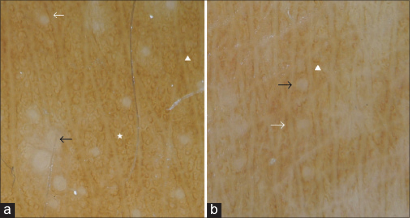
A 17-year-old girl presented with brownish macules arranged in a whorled and linear pattern along the lines of Blaschko on the trunk and limbs [Figure - 1]a and [Figure - 1]b. These asymptomatic lesions had initially appeared over her trunk when she was two months old, and gradually progressed to involve both upper and lower limbs by the time she turned one. There was no history of previous vesiculo-bullous or inflammatory lesions and similar lesions were not present in any family member. There were no abnormalities of the mucosae or skin appendages. The girl had normal developmental milestones and systemic examination was within normal limits On dermoscopy, the whorled patches on the trunk showed numerous brownish rings and curved lines and a few brownish streak-like lines [Figure - 2]a, while the linear patches on the legs showed short brownish streak-like lines arranged in a parallel manner and a few curved brownish lines [Figure - 2]b. Another dermoscopic finding present in both linear and whorled lesions consisted of focally distributed hypopigmented dots corresponding to perifollicular areas [Figure - 2]a and [Figure - 2]b.
 |
| Figure 1: Brownish macules arranged in a whorled and linear pattern along the lines of Blaschko on the trunk (a) and limbs (b) |
 |
| Figure 2: (a) Dermoscopic view shows numerous brownish rings (white star) and curved lines (white arrowhead) and a few brownish streak-like lines (white arrow) on the whorled patches of the trunk and (b) short brownish streak-like lines arranged in a parallel way (white arrow) and a few curved brownish lines (white arrowhead) on the linear patches of the legs. Focally distributed hypopigmented dots (black arrow), corresponding to perifollicular areas, are also present in both whorled (a) and linear (b) lesions (×10) |
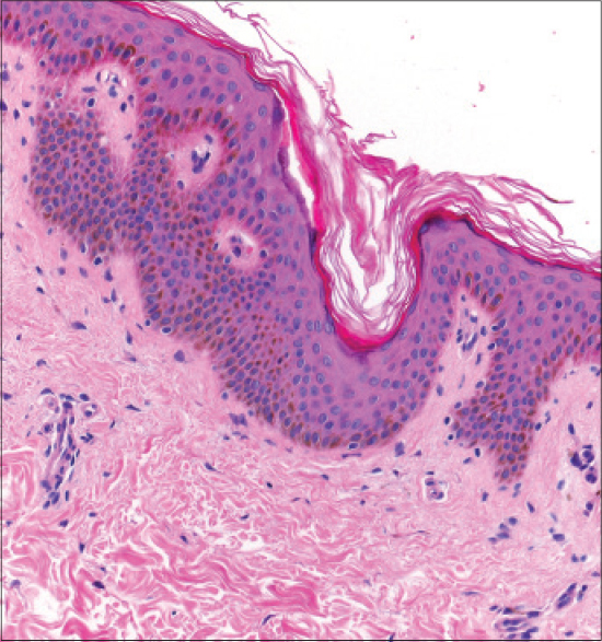
A skin biopsy from a lesion on her left thigh confirmed the clinical diagnosis of linear and whorled nevoid hypermelanosis by revealing increased basal layer pigmentation without melanocytosis; there was no pigment incontinence in the dermis [Figure - 3]. Genetic studies to evaluate the presence of chimerism or mosaicism were not done as the girl's parents refused. The patient was reassured and explained about the benign nature of the disorder; no treatment was given.
 |
| Figure 3: Increased basal layer pigmentation without melanocytosis; there is no pigment incontinence in the dermis (H and E, ×200) |
Dermoscopic features of linear and whorled nevoid hypermelanosis have been described in two previous instances and the findings were found to be discordant.[3],[4] Naveen and Reshme reported a “net like” pattern of pigmentation noted in both linear and whorled lesions while Ertam et al. described a “parallel” pattern which consisted of linear or circular arrangements of parallel brown streaks with an overall alignment along the lines of Blaschko.[3],[4] In particular, on the epigastric region, due to the whorled-like configuration of the lesions, the authors found partially curved, circular and/or linear parallel pigmented streaks in a parallel pattern while on the popliteal region, the same curved and/or linear streak-like pigmentations were vertical to skin profiles, but parallel to each other.[4] The dermoscopic features described in our case are reminiscent of the “parallel” pattern. Moreover, the predominance of linear streaks on the linear patches on the legs, and rings/curved lines on the whorled lesions of the back may be justified by the well-known regional variations in morphology of Blaschko's lines.[4] However, our patient showed one additional previously unreported dermoscopic feature, namely, dotted perifollicular hypopigmentation. Interestingly, these dots did not involve all the follicular ostia in the lesional areas which is in keeping with the mosaic nature of the disorder.[3]
In conclusion, our case emphasizes that linear and whorled nevoid hypermelanosis may be dermoscopically heterogeneous, displaying some individual variations. It also confirms that dermoscopy might be useful in differentiating this disorder from its prime differential diagnosis, namely, incontinentia pigmenti, which is characterized by blue-gray dots histologically corresponding to pigmentary incontinence.[3]
Financial support and sponsorship
Nil.
Conflicts of interest
There are no conflicts of interest.
| 1. |
Metta AK, Ramachandra S, Sadath N, Manupati S. Linear and whorled nevoid hypermelanosis in three successive generations. Indian J Dermatol Venereol Leprol 2011;77:403.
[Google Scholar]
|
| 2. |
Di Lernia V. Linear and whorled hypermelanosis. Pediatr Dermatol 2007;24:205-10.
[Google Scholar]
|
| 3. |
Naveen KN, Reshme P. Linear and whorled nevoid hypermelanosis with dermatoscopic features. Dermatol Online J 2014;20:13.
[Google Scholar]
|
| 4. |
Ertam I, Turk BG, Urkmez A, Kazandi A, Ozdemir F. Linear and whorled nevoid hypermelanosis: Dermatoscopic features. J Am Acad Dermatol 2009;60:328-31.
[Google Scholar]
|
Fulltext Views
5,027
PDF downloads
2,281


