Translate this page into:
Linear eccrine angiomatous hamartoma
Corresponding author: Dr. Ruovinuo Theunuo, Department of Dermatology, ESIC Medical College Faridabad, Haryana, India. truovinuo@gmail.com
-
Received: ,
Accepted: ,
How to cite this article: Theunuo R, Ramesh V, Passi S, Uikey D. Linear eccrine angiomatous hamartoma. Indian J Dermatol Venereol Leprol doi: 10.25259/IJDVL_527_2023
Dear Editor,
A 7-year-old boy presented with unilateral, multiple raised lesions over the left lower leg of 4-years duration. On examination, there were multiple, discrete, brown-black, firm, well-defined plaques ranging from 1.5 to 2cm in diameter, arranged in a linear pattern starting from the anterolateral aspect of the thigh down to the ankle, with some of the lesions showing hemorrhagic crusts and hypertrichosis [Figures 1a and 1b]. On the calf, one lesion had a nodular and tender subcutaneous component better felt than seen. There was no complaint of hyperhidrosis. Dermoscopy revealed multiple yellow nodules, dotted vessels, and red lacunae [Figure 2]. Histopathology showed mild hyperkeratosis with dilated cavernous vessels in the papillary dermis extending into the upper dermis. Mature hair follicles were seen in the subcutis while eccrine sweat glands were grouped near the follicle and scattered in the dermis [Figures 3a, 3b and 3c]. Hence, a diagnosis of linear eccrine angiomatous hamartoma was made. The parents were counseled regarding the possibility of spontaneous resolution as the child was uncooperative for minor surgical procedure.

- Linear plaques over left leg
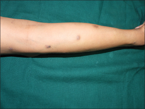
- Plaques showing skin-colored and angiomatous papules with hypertrichosis
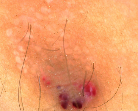
- Dermoscopy (polarized mode)-shows multiple yellow globules, dotted vessels, red and purple lacunae. Hypertrichosis can be seen
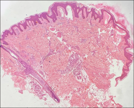
- Cavernous vessels in the papillary dermis, eccrine glands, and mature hair follicles in the dermis (H&E, x40)
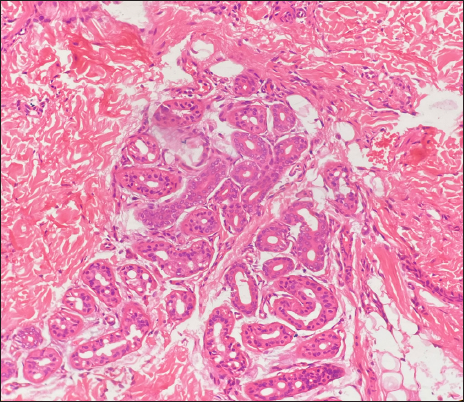
- Collection of eccrine glands in the dermis (H&E, x200)
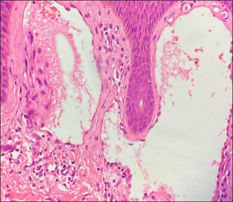
- Cavernous vessels in the papillary dermis (H&E, x400)
Eccrine angiomatous hamartoma (EAH) is an uncommon, benign condition characterized by the proliferation of eccrine glands and capillary channels. The condition was first described by Lotzbeck in 1859 1 , and the term was coined by Hyman et al in 1968. Although the etiology is unclear, it is thought to be caused by defective biochemical interactions between the epithelium and the mesenchyme.
It usually presents at birth or during childhood as a solitary skin-colored, reddish, violaceous, or blue-brown nodule or plaque and less commonly as a macule or patch. Common sites of involvement include the limbs, particularly the palms, and soles, followed by the trunk, head, and neck. Rarely, a callus-like presentations has been reported. 2 The lesions grow in proportion to the growth of the individual. A few cases with onset in adulthood have been reported, and they are proposed to be linked with recurrent trauma. While lesions can be asymptomatic, pain and hyperhidrosis are present in 42% and 32% of patients. Additionally, hypertrichosis and local rise in temperature are found in some cases. Some authors believe that symptoms can worsen in puberty and pregnancy attributable to hormonal influences.
Dermoscopy shows the popcorn nodular pattern over a background of erythema and linear and arborizing blood vessels, 3 somewhat like our observation. On histopathology, we saw cavernous and dilated vessels in the dermis, increased eccrine sweat glands, and mature hair follicles. Other components described are lymphatic vessels in the deep dermis with fat, mucin, apocrine or neural components. Pain is due to small nerves penetrating the lesion.
Of around 100 documented cases, there are several reports of multiple lesions and segmental distribution, but we found only three reports of linear eccrine angiomatous hamartoma. One had linear lesions in the inguinal fold 4 , while another had linearly arranged macules from the tip of the middle finger to the palm. 5 Patterson et al reported a case with linear erythematous plaques on the posterior aspect of the leg. 6 The condition has also been described to co-exist with verrucous epidermal nevus and nevus sebaceous as part of linear ectodermal cutaneous hamartoma in a single patient. 7
Differential diagnosis includes eccrine nevus, eccrine poroma, glomus tumor, tufted angioma, smooth muscle hamartoma, and blue rubber bleb nevus. Definitive treatment is surgical resection. Other modalities for symptomatic patients include pulsed dye laser, Nd: YAG laser, and sclerotherapy. Botulinum toxin injection can be given if there is hyperhidrosis. Spontaneous regression has also been observed.
Declaration of patient consent
The authors certify that they have obtained all appropriate patient consent.
Financial support and sponsorship
Nil.
Conflict of interest
There is no conflict of interest.
References
- Ein Fall von Schweissdruesengeschwulst an der Wauge. Virchow Arch Pathol Anat. 1859;16:160.
- [Google Scholar]
- Eccrine angiomatous hamartoma: a review of ten cases. Ann Dermatol. 2013;25:208-12.
- [CrossRef] [PubMed] [PubMed Central] [Google Scholar]
- Dermoscopy of eccrine angiomatous hamartoma: the popcorn pattern. JAAD Case Rep. 2018;4:165-167.
- [CrossRef] [PubMed] [Google Scholar]
- Eccrine angiomatous harmartoma in an infant. Pediatr Dermatol. 2006;23:365-368.
- [CrossRef] [PubMed] [Google Scholar]
- Hamartoma angiomatoso ecrino: presentación de dos casos [Eccrine angiomatous hamartoma: a report of 2 cases] Actas Dermosifiliogr. 2011;102:289-292.
- [CrossRef] [PubMed] [Google Scholar]
- Eccrine Angiomatous Hamartoma: A Clinicopathologic Review of 18 Cases. Am J Dermatopathol. 2016;38:413-7.
- [CrossRef] [PubMed] [Google Scholar]
- Linear ectodermal cutaneous hamartoma. Int J Dermatol. 2003;42:376-9.
- [CrossRef] [PubMed] [Google Scholar]


