Generic selectors
Exact matches only
Search in title
Search in content
Post Type Selectors
Search in posts
Search in pages
Filter by Categories
15th National Conference of the IAOMFP, Chennai, 2006
Abstract
Abstracts from current literature
Acne in India: Guidelines for management - IAA Consensus Document
Addendum
Announcement
Art & Psychiatry
Article
Articles
Association Activities
Association Notes
Award Article
Book Review
Brief Report
Case Analysis
Case Letter
Case Letters
Case Notes
Case Report
Case Reports
Clinical and Laboratory Investigations
Clinical Article
Clinical Studies
Clinical Study
Commentary
Conference Oration
Conference Summary
Continuing Medical Education
Correspondence
Corrigendum
Cosmetic Dermatology
Cosmetology
Current Best Evidence
Current Issue
Current View
Derma Quest
Dermato Surgery
Dermatopathology
Dermatosurgery Specials
Dispensing Pearl
Do you know?
Drug Dialogues
e-IJDVL
Editor Speaks
Editorial
Editorial Remarks
Editorial Report
Editorial Report - 2007
Editorial report for 2004-2005
Errata
Erratum
Focus
Fourth All India Conference Programme
From Our Book Shelf
From the Desk of Chief Editor
General
Get Set for Net
Get set for the net
Guest Article
Guest Editorial
History
How I Manage?
IADVL Announcement
IADVL Announcements
IJDVL Awards
IJDVL AWARDS 2015
IJDVL Awards 2018
IJDVL Awards 2019
IJDVL Awards 2020
IJDVL International Awards 2018
Images in Clinical Practice
In Memorium
Inaugural Address
Index
Knowledge From World Contemporaries
Leprosy Section
Letter in Response to Previous Publication
Letter to Editor
Letter to the Editor
Letter to the Editor - Case Letter
Letter to the Editor - Letter in Response to Published Article
LETTER TO THE EDITOR - LETTERS IN RESPONSE TO PUBLISHED ARTICLES
Letter to the Editor - Observation Letter
Letter to the Editor - Study Letter
Letter to the Editor - Therapy Letter
Letter to the Editor: Articles in Response to Previously Published Articles
Letters in Response to Previous Publication
Letters to the Editor
Letters to the Editor - Letter in Response to Previously Published Articles
Letters to the Editor: Case Letters
Letters to the Editor: Letters in Response to Previously Published Articles
Medicolegal Window
Messages
Miscellaneous Letter
Musings
Net Case
Net case report
Net Image
Net Images
Net Letter
Net Quiz
Net Study
New Preparations
News
News & Views
Obituary
Observation Letter
Observation Letters
Oration
Original Article
ORIGINAL CONTRIBUTION
Original Contributions
Pattern of Skin Diseases
Pearls
Pediatric Dermatology
Pediatric Rounds
Perspective
Presedential Address
Presidential Address
Presidents Remarks
Quiz
Recommendations
Regret
Report
Report of chief editor
Report of Hon : Treasurer IADVL
Report of Hon. General Secretary IADVL
Research Methdology
Research Methodology
Resident page
Resident's Page
Resident’s Page
Residents' Corner
Residents' Corner
Residents' Page
Retraction
Review
Review Article
Review Articles
Reviewers 2022
Revision Corner
Self Assessment Programme
SEMINAR
Seminar: Chronic Arsenicosis in India
Seminar: HIV Infection
Short Communication
Short Communications
Short Report
Snippets
Special Article
Specialty Interface
Studies
Study Letter
Study Letters
Supplement-Photoprotection
Supplement-Psoriasis
Symposium - Contact Dermatitis
Symposium - Lasers
Symposium - Pediatric Dermatoses
Symposium - Psoriasis
Symposium - Vesicobullous Disorders
SYMPOSIUM - VITILIGO
Symposium Aesthetic Surgery
Symposium Dermatopathology
Symposium-Hair Disorders
Symposium-Nails Part I
Symposium-Nails-Part II
Systematic Review and Meta-Analysis
Systematic Reviews and Meta-analyses
Systematic Reviews and Meta-analysis
Tables
Technology
Therapeutic Guideline-IADVL
Therapeutic Guidelines
Therapeutic Guidelines - IADVL
Therapeutics
Therapy
Therapy Letter
Therapy Letters
View Point
Viewpoint
What’s new in Dermatology
View/Download PDF

Translate this page into:

Letter to the Editor
2010:76:6;707-709
doi: 10.4103/0378-6323.72452
PMID: 21079323

Localization of T-cell subsets in cutaneous lichen planus: An insight into pathogenetic mechanism

Safia Rana1 , Ruchika Gupta2 , Sompal Singh1 , Sandip Mohanty3 , Kusum Gupta1 , Madhur Kudesia1
1 Department of Pathology, Hindu Rao Hospital, Delhi, India
2 Department of Pathology, All India Institute of Medical Sciences, Delhi, India
3 Department of Skin V.D., Hindu Rao Hospital, Delhi, India

Correspondence Address:
Ruchika Gupta
Department of Pathology, All India Institute of Medical Sciences, Ansari Nagar, New Delhi - 10029
India
How to cite this article:
Rana S, Gupta R, Singh S, Mohanty S, Gupta K, Kudesia M. Localization of T-cell subsets in cutaneous lichen planus: An insight into pathogenetic mechanism. Indian J Dermatol Venereol Leprol 2010;76:707-709
Copyright: (C)2010 Indian Journal of Dermatology, Venereology, and Leprology

Sir,

We would like to report the results of our evaluation of T-cell subsets in cutaneous lichen planus (LP). For the study, 20 cases of cutaneous LP over a one-year period (2007-2008) were included and diagnosis confirmed by a review of the clinical and histopathological features. Patients with features suggestive of systemic lupus erythematosus, human immunodeficiency virus infection or other chronic medical illnesses were excluded. Immunohistochemistry for T-cell helper subset (CD4, BioGenex, USA) and T-cell cytotoxic/ suppressor subset (CD8, BioGenex, USA) was performed on serial sections using the immunoperoxidase technique. The biopsies were examined for inflammatory cells at two locations - dermo-epidermal junction and upper reticular dermis (perivascular locations). Quantification of CD4 and CD8 T cells was performed by two independent observers at X200 magnification. A mean of the observations were taken and the relative proportion (percentage) of CD4 and CD8 subsets of T cells at each location was calculated. Non-parametric test was used to find the significance of difference in T cell subsets at the two locations.

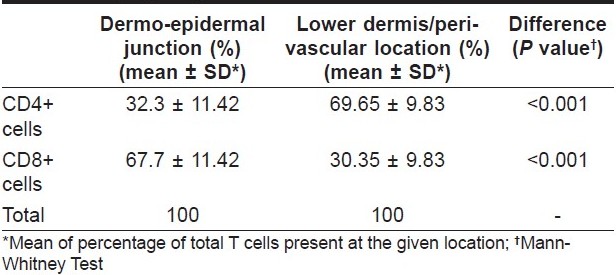
The mean age of our cases was 36.95 ± 16.32 years with slight predominance of females (male: female 1:1.5). Skin biopsies in all cases showed the characteristic histopathological features of lichen planus. At the dermo-epidermal location, the percentage of CD4+ cells ranged from 10 to 50% of total number of T cells, with a mean of 32.3% of cells. On the other hand, CD8+ T cells [Figure - 1]a, b ranged from 52 to 90%, with a mean of 67.7% of cells. In contrast, the peri-vascular location showed preponderance of CD4+ cells [Figure - 1]c ranging from 50 to 88% (mean 69.65%) in the sparse number of T cells present. The CD8+ T cells ranged from 12 to 50%, with a mean of 30.35% of cells.

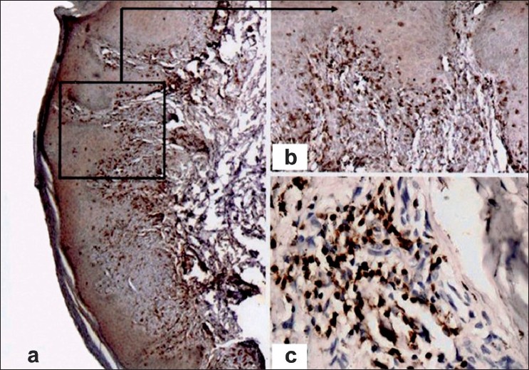
Figure 1: Photomicrograph showing immunohistochemical staining in cutaneous lichen planus with (a, ×100) CD8 demonstrating CD8-positive cells at the dermo-epidermal junction, (b, ×200) higher power view demonstrating the CD8-positive cells, (c, ×200) CD4 staining showing CD4-positive cells in the perivascular location

The relative percentages of CD 4+ and CD 8+ T cells at the dermo-epidermal junction and in lower dermis are tabulated in [Table - 1]. The difference in percentage of CD 4+ T cells between dermoepidermal junction and perivascular location was statistically significant (Mann-Whitney test, p <0.001). Similar results were obtained for CD8+ T cells, as shown in [Table - 1].

Table 1: Localization of CD4+ and CD8+ subtypes of T lymphocytes in cutaneous lichen planus

LP is a chronic muco-cutaneous inflammatory disease with typical histopathological changes of compact orthokeratosis with hypergranulosis, irregular acanthosis, basal cell damage and a band-like infiltrate of lymphocytes. [1] Elaborate studies have been performed in cases of oral LP to analyze the subsets of T lymphocytes. The majority of subepithelial and intraepithelial inflammatory cells in oral LP are CD8+ T cells. [2],[3] Activated CD8+ T cells trigger basal cell apoptosis through perforin/ granzyme or Fas/ Fas ligand pathways. [2] At the same time, most of the lymphocytes in lamina propria of oral LP are CD4+ helper T cells. [4] In cutaneous LP also, the inflammatory cells have been shown to be T lymphocytes with majority expressing HLA-DR antigen and some showing expression of IL-2 receptor. [5] Earlier analysis of the various subtypes of T cells has yielded contradictory results, with some authors demonstrating predominance of helper-inducer CD4+ T cells and others reporting suppressor-cytotoxic T lymphocytes as the main subset present in the infiltrate. [4],[6] We found the presence of CD8+ effector T cells as well as CD4+ helper T cells in cutaneous LP. The present study supports the primary role of CD8+ cytotoxic T cells in keratinocyte damage, since the dermo-epidermal infiltrate was seen to be composed predominantly of CD8+ T cells. The perivascular localization of CD4+ T cells is an indicator of the possible assistance offered to the cytotoxic CD8+ cells by secretion of Th1 cytokines. Keratinocyte damage by CD8+ T cells would lead to basement membrane disruption, paving way for the influx of more CD8+ T cells and further basal cell damage resulting in a vicious cycle leading to chronicity of the disease. Our results on cutaneous lichen planus are similar to those postulated for oral LP. [2],[3] The predominance of T cells in the inflammatory infiltrate, as seen in our study, also excludes a primary role of humoral immune mechanism in the pathogenesis of cutaneous LP. With the advent of directed immunotherapy, a detailed study of the pathogenetic mechanisms would provide further refinement of the therapeutic strategies for patients with cutaneous lichen planus. The results of the present study require to be confirmed in further studies.

In conclusion, cutaneous lichen planus is a T-cell mediated inflammatory disease with pathogenetic role of both CD8+ cytotoxic T cells and CD4+ helper T cells. There is a predominance of cytotoxic T cells in the dermo-epidermal infiltrate responsible for keratinocyte damage. Helper/ inducer CD4+ T cells are found in the perivascular dermal infiltrate, which may be assisting the cytotoxic cells in the keratinocyte damage.

References
1.
Ellis FA. Histopathology of lichen planus based on analysis of one hundred biopsies. J Invest Dermatol 1967;48:143-8.
[Google Scholar]
2.
Sugerman PB, Savage NW, Walsh LJ, Zhao ZZ, Zhou XJ, Khan A, et al. The pathogenesis of oral lichen planus. Crit Rev Oral Biol Med 2002;13:350-65.
[Google Scholar]
3.
Iijima W, Ohtani H, Nakayama T, Sugawara Y, Sato E, Nagura H, et al. infiltrating CD8+ T Cells in oral lichen planus predominantly express CCR5 and CXCR3 and carry respective chemokine ligands RANTES/CCL5 and IP-10/CXCL10 in their cytolytic granules: a potential self-recruiting mechanism. Am J Pathol 2003;163:261-8.
[Google Scholar]
4.
Ishii T. Immunohistochemical demonstration of T cell subsets and accessory cells in oral lichen planus. J Oral Pathol 1987;16:356-61.
[Google Scholar]
5.
Sundquist KG, Wanger L. Expression of lymphocyte activation markers in benign cutaneous T-cell infiltrate: discoid lupus erythematosus versus lichen ruber planus. Acta Derm Venereol (Stockh) 1989;69:292-5.
[Google Scholar]
6.
Buechner SA. T-cell subsets and macrophages in lichen planus. Dermatologica 1984;169:325-9.
[Google Scholar]

Fulltext Views
2,396

PDF downloads
950
Show Sections