Translate this page into:
Malignant blue nevus
Correspondence Address:
M Jayaraman
222 R.K. Mutt Road, Mylapore, Madras - 600 004
India
| How to cite this article: Krishnan S, Yesudian DP, Jayaraman M, Janaki V R, Yesudian P. Malignant blue nevus. Indian J Dermatol Venereol Leprol 1997;63:183-185 |
Abstract
Blue nevus represents an aberrant collection of functioning benign dermal melanocytes. Its malignant degeneration is rare and is regarded as a form of malignant melanoma. We report a case of 35-year old male with this rare condition whose primary lesion over left foot ulcerated and patient later succumbed to multiple metastases. |
 |
 |
 |
 |
 |
Blue nevus is an area of blue or black dermal pigmentation produced by aberrant collection of functioning benign melanocytes. Blue nevus of skin was described in 1906 by Tieche.[1] Since then it has been reported to occur at other body sites. Three types of blue nevi have been recognised on the skin-common, cellular and combined.[2] Malignant degeneration of these nevi is extremely rare and is regarded as a rare form of malignant melanoma. We report a case of this rare condition.
Case Report
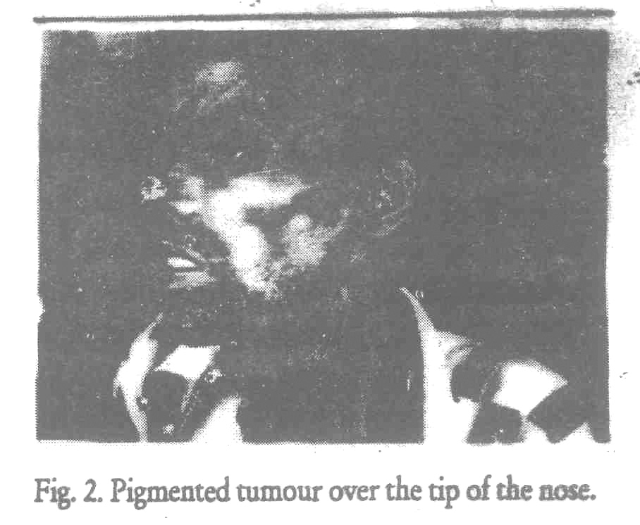
The patient, 35-year-old male, had at birth a circumscribed slightly raised, bluish lesion about 1.5 cm in diameter over the dorsum of the left foot along the medial margin. The lesion had remained silent for 35 years. Recently, for the past 6 months, patient had noticed an increase in size of the lesion along with ulceration [Figure - 1]. Over the past 3 months he had developed bluish pigmented tumor over the tip of the nose [Figure - 2] and multiple bluish black nodules over the thigh. He had associated loss of appetite and weight. On examination he was anemic, emaciated and had multiple enlarged inguinal lymphnodes, which were discrete and firm to hard in consistency.
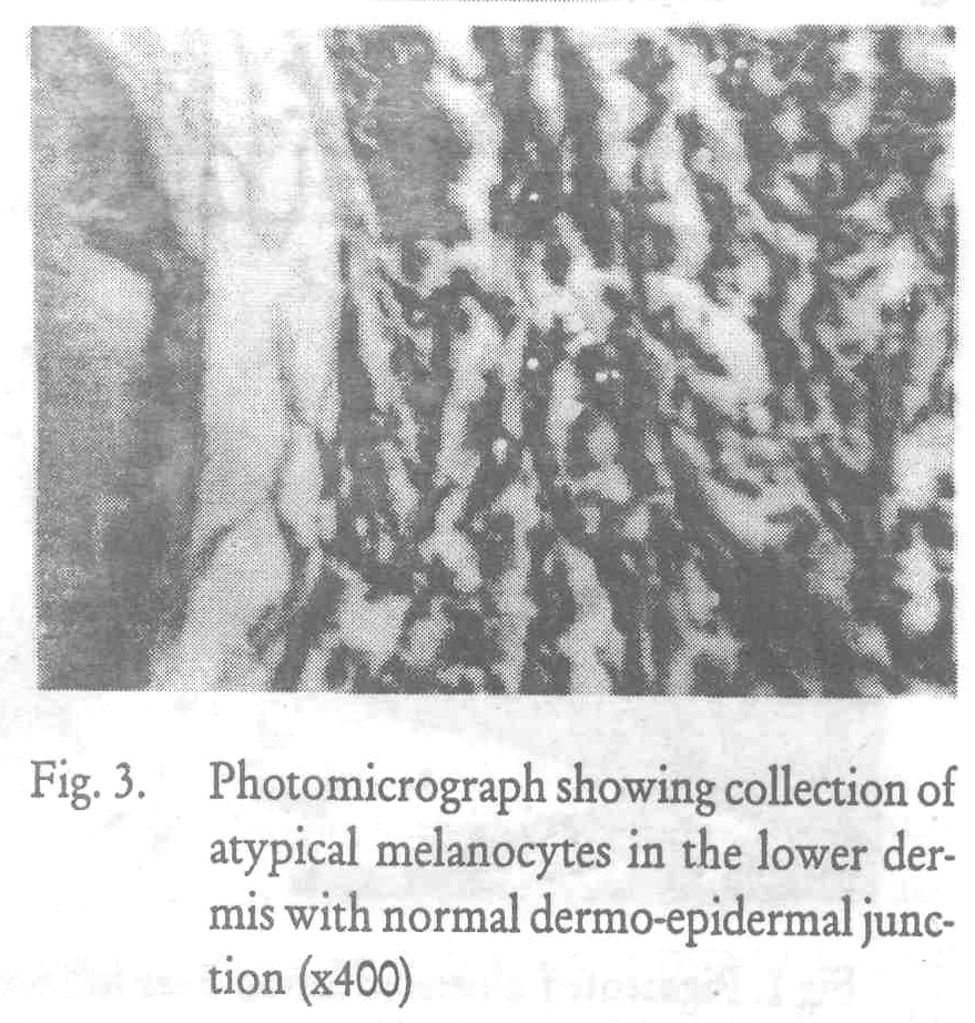
Biopsy of one of the pigmented nodules of the thigh revealed dense collection of dendritic pigmented cells in the mid and lower dermis. Some of the cells had hyperchromatic bizzare nuclei. A few atypical mitoses were also seen. Overlying upper dermis and epidermis were normal, thus giving a diagnosis of malignant blue nevus [Figure - 3]. Radiography of the chest and bones revealed multiple metastases. Lymph node biopsy was not done. Patient was put on chemotherapy, but died 3 months later.
Discussion
Malignant blue nevus is a rare form of malignant melanoma. It may arise in a blue nevus, nevus of Ota or may be malignant from start. Those arising in a blue nevus occur only in cellular type. The most common site for its occurrence is scalp, even though blue nevi most commonly occur on the dorsum of the hands and feet. Most of the malignant blue nevi at the time of diagnosis are more than 3 cm. in diameter.[3] Metastasis occurs rapidly and lymphnodes are involved invariably. Here we have to be careful to exclude "benign metastasis" where cases of benign cellular blue nevi are associated with regional lymphnode collection of cells histologically similar to the primary lesion.[4] In general, histopathology of the metastases resembles those of the primary lesion except for the difference in the degree of necrosis / cellular pleomorphism.
Our patient had ulceration of the blue nevus which is very rare and has been reported only in 2 cases previously. [5,6] The rapid progression of the disease with multiple metastases early in the course and lack of response to treatment makes it essential to diagnose the condition at the outset. The common occurrence of blue nevus makes it inappropriate to excercise all of them prophylactically, but it would be advisable to remove those greater than 2cm. in diameter.
| 1. |
Tieche M. Uber benigne melanome der hant. "blue naevi", Arch Pathol Anat, 1906;186:212-229.
[Google Scholar]
|
| 2. |
Lever WF, Lever GS. Benign melanocytic tumors and malignant melanoma, In : Histopathology of Skin, 7th edn, JB. Lippincott, Philadelphia 1990;19:712-722.
[Google Scholar]
|
| 3. |
Goldenhersh MS, Savin RC, Barnhill RL, et al. Malignant blue nevus. A case report and review of the literature, J Am Acad Dermatol 1988;19:712-722.
[Google Scholar]
|
| 4. |
Allen AC, Spitzs. Malignant melanoma, Cancer 1953;6:1-45.
[Google Scholar]
|
| 5. |
Jadassohn W, Franceschetti A, Quelques GM. Observations cliniques concernant la pigmentation duderme, Dermatologica 1954;108:225-234.
[Google Scholar]
|
| 6. |
Herzberg JJ, Klein UE. Blue nevus mit solitar metastasen in lunge and nebennieren, Arch Klin Exp Dermatol 1961;212:158-172.
[Google Scholar]
|


