Translate this page into:
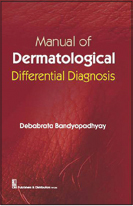
Manual of Dermatological Differential Diagnosis
Correspondence Address:
M Manjunath Shenoy
Department of Dermatology, Yenepoya Medical College, Deralakatte, Mangalore, Karnataka
India
| How to cite this article: Shenoy M M. Manual of Dermatological Differential Diagnosis. Indian J Dermatol Venereol Leprol 2016;82:595 |
Author: Debabrata Bandyopadhyay
Publisher: CBS Publishers and Distributors Pvt. Ltd., New Delhi, India
ISBN: 978-81-239-2942-2
Edition: First Edition, 2016
Pages: 307
Price: Rs. 350/-

Differential diagnosis in medical science refers to distinguishing a disease or a symptom or a sign of a particular clinical condition from others that present in a similar fashion. It is a systematic way in which clinicians put forward various diagnostic options and then exclude them one by one using their clinical acumen. In dermatology, most clinical signs of diseases are visible and may indicate the possibility of an internal disease. This gives us an array of differential diagnosis, making it one of the most challenging subspecialties in medicine. Manuals on differential diagnosis become an essential addition to the existing handbooks and, in my opinion, are value-added services provided by the author to this expanding field of dermatology.
Critical analysis of this book reveals that it is typically a manual that revolves around the morphology and distribution of cutaneous lesions. There is a brief yet crystal clear description of the morphology, followed by a list of clinical differentials. For example, guttate leukoderma is described as well-demarcated, small, drop-shaped, hypopigmented or depigmented macules and this is followed by a list of eight differential diagnoses. Arrangements (preferred term being configuration) of the lesions such as grouped, serpiginous, figurate and many others have been adequately covered with simple descriptions and a good number of differentials. Some of the entities discussed here such as “hyperhidrosis” though appear like a misfit under the heading of morphology and arrangements and may have been included in view of unavailability of an appropriate section for this topic.
The book has a very good collection of signs that are pertinent to the field of dermatology. Out of the total 196, most of them are truly named signs and others are similes; for example, mulberry molar, moon facies. This is a large collection and it will hence be a delight for the examiners and examinees. Another interesting feature is the section on dermatoses and drugs, in which cutaneous, mucosal, hair and nail changes caused by drugs are described. A brief description of some of the entities such as baboon syndrome or photo recall dermatitis could have been given. Rest of the book is dedicated to the diagnostic features of dermatoses. Common and rare diseases are covered in this section with brief descriptions of the disease with some of the best clinical images. Where necessary, classification of the diseases has also been dealt with. This section can be good reading for students on the previous day of the examination.
A strong feature of the book is that in spite of lots of information yet, it continues to remain a handbook. I congratulate the author and the publisher for bringing out another useful book and recommend this book to students, teachers and clinical dermatologists.
Fulltext Views
4,648
PDF downloads
2,000


