Translate this page into:
Median raphe cyst of penis
Correspondence Address:
Deepali Chandrakant Tarate
Ashtavinayak Society, Flat No. 1801, Ekta Nagar, Kandivali (West), Mumbai - 400 067, Maharashtra
India
| How to cite this article: Tarate DC, Tambe SA, Nayak CS. Median raphe cyst of penis. Indian J Dermatol Venereol Leprol 2018;84:373 |
Sir,
Median raphe cyst is a rare, benign congenital cyst presenting most commonly on the ventral aspect of penile shaft. These lesions present mostly in young men, whereas some cases in children have also been reported. It was first described by Mermet in 1895[1] and by Lantin and Thompson in 1956.[2] It usually presents anywhere along the median raphe, i.e., in the midline from the urethral meatus extending ventrally to the anus.[3],[4]
Two married men aged 26 years and 30 years presented with a round swelling on the penis for 8 and 10 years, respectively. The swelling gradually increased in size and was associated with pain and discomfort during sexual intercourse. There was no history of trauma, infection or other relevant clinical history.
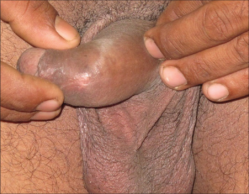
Cutaneous examination of the first patient revealed a single well-defined non-tender smooth globular cystic lesion measuring 1 cm × 1 cm on the ventral aspect of prepuce with overlying normal skin [Figure - 1]a. Transillumination test was positive. Cutaneous examination in the second patient revealed a single well-defined non-tender smooth globular cystic lesion measuring 0.5 cm × 0.5 cm on the ventral aspect of glans penis in a parameatal location [Figure - 1]b. Excision biopsy was undertaken in both the patients.
 |
| Figure 1 |
Histopathology of the lesions showed compact stratum corneum overlying acanthotic epidermis with evidence of a disrupted cyst in the dermis showing an epithelial lining of stratified squamous and columnar (mixed) cells [Figure - 2]a and [Figure - 2]b. The postoperative follow-up revealed no recurrence in the first patient at 1-year follow-up and the second patient was lost to follow-up [Figure - 3].
 |
| Figure 2: (a) Evidence of a disrupted cyst in the dermis. (b) Cyst wall showing epithelial lining of stratified squamous and columnar cells (mixed), (H and E, ×400) |
 |
| Figure 3: Follow-up of first patient at 1 year |
Median raphe cysts are rarely reported since they are usually asymptomatic. They are not noticed during childhood and usually present during adulthood with difficulty in micturition and difficulty in having sexual intercourse. The most common location of such cysts is the ventral aspect of penile shaft and in the parameatal position.[5] These cysts were previously reported as genito-perineal cyst of the median raphe, mucous cyst of the penis and parameatal cyst.[6]
Various theories have been considered as the exact pathogenesis is not known. According to Lantin and Thompson, median raphe cyst occurs in the process of separation of the foreskin from the glans penis,[2] Littre theorizes its occurrence due to the presence of ectopic periurethral glands.[4] The tissue trapping theory attributes its occurrence either due to a defective fusion of the urethral folds or an anomalous outgrowth of the epithelium that becomes sequestrated and independent after the primary closure of the median raphe. This theory also explains the different types of epithelial lining it has: (1) pseudostratified columnar epithelium (trapping of proximal urethral cells), (2) squamous cell epithelium (trapping of distal urethral cells), (3) glandular epithelium (trapping of periurethral glands) and (4) the mixed type.
Treatment options include simple aspiration, wide local excision and marsupialization or deroofing for deeply located large cysts. Postoperative complications such as urethrocutaneous fistula, gaping sinuses and recurrences are seen with deroofing and aspiration of the cyst.[5] Complete local excision with primary closure is the treatment of choice.
Financial support and sponsorship
Nil.
Conflicts of interest
There are no conflicts of interest.
| 1. |
Mermet P. Congenital cysts of the genitoperineal raphe. Rev Chir 1895;15:382-435.
[Google Scholar]
|
| 2. |
Lantin PM, Thompson IM. Parameatal cysts of the glans penis. J Urol 1956;76:753-5.
[Google Scholar]
|
| 3. |
Nagore E, Sánchez-Motilla JM, Febrer MI, Aliaga A. Median raphe cysts of the penis: A report of five cases. Pediatr Dermatol 1998;15:191-3.
[Google Scholar]
|
| 4. |
Kirkham N. Tumors and cysts of the epidermis. In: Elder D, Elenitsas R, Jasorsky C, Jonhson B, editors. Lever's Histopathology of Lever. 8th ed. Philadelphia: Lippincott-Raven; 1997. p. 685-746.
[Google Scholar]
|
| 5. |
Shao IH, Chen TD, Shao HT, Chen HW. Male median raphe cysts: Serial retrospective analysis and histopathological classification. Diagn Pathol 2012;7:121.
[Google Scholar]
|
| 6. |
Otsuka T, Ueda Y, Terauchi M, Kinoshita Y. Median raphe (parameatal) cysts of the penis. J Urol 1998;159:1918-20.
[Google Scholar]
|
Fulltext Views
5,239
PDF downloads
3,146


