Translate this page into:
Medicopsis romeroi: A causative agent of subcutaneous phaeohyphomycosis in a diabetic patient
Corresponding author: Dr. Seema Rani, Department of Dermatology, Lady Hardinge Medical College & Smt S. K. Hospital, Delhi, India. drseemashekhar@gmail.com
-
Received: ,
Accepted: ,
How to cite this article: Kudari SM, Rani S, Mendiratta V, Sonker S, Rawat D, Xess I, et al. Medicopsis romeroi: A causative agent of subcutaneous phaeohyphomycosis in a diabetic patient. Indian J Dermatol Venereol Leprol. doi: 10.25259/IJDVL_675_2023
Dear Editor,
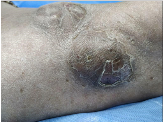
A 66-year-old woman, a homemaker, presented with a painless cystic swelling on her left foot of eight months duration. She was diabetic for the past five years. There was no history of preceding trauma and pus/grain discharge from the lesion. Examination revealed a well-defined, soft-to-firm, non-tender nodulo-cystic swelling of 3 × 3 cm, on the medial side of the plantar surface of her left foot [Figure 1]. Another lesion of similar morphology with surface lobulations was present adjacent to it. We kept a differential of deep fungal infection, botryomycosis, and tubercular abscess. Direct microscopic examination with 10% potassium hydroxide and fine needle aspiration cytology showed septate fungal hyphae. Histopathology revealed dense granulomatous inflammatory infiltrate along with occasional giant cells and slender hyphae with occasional spores in the deep dermis, [Figure 2] which were periodic acid-schiff (PAS) and Grocott methenamine silver stain positive [Figure 3]. The aspirated content was sent for bacteriological and fungal culture (Sabouraud dextrose agar with chloramphenicol). The macroscopic aspect of the culture was cottony, greyish-white, rugose on the obverse side and black pigmentation on the inverse side. Molecular identification was performed by polymerase chain reaction amplification and sequencing was done from the culture growth. The internal transcribed spacer (ITS) 1 and ITS2 regions and the 5.8S ribosomal DNA region of the fungi were amplified by using universal primers ITS1 and ITS4. The sequence was submitted to GenBank with accession number OP764412. The clinical isolate was Medicopsis romeroi. Lesional ultrasonography showed a well-defined multilobulated cystic lesion in the subcutaneous plane and a few internal hyperechoic foci with posterior acoustic shadowing without any internal vascularity. Magnetic resonance imaging of the left foot revealed well-defined multilobulated cystic lesions with thick and nodular-enhancing septa involving the subcutaneous plane without involvement in muscles, joint cavity, or bony destruction. Routine biochemical investigations were within the normal limits. Her glycated haemoglobin level was 6.5. Lesions were surgically excised and itraconazole (200 mg) was given for 12 weeks but later stopped by the patient herself. The patient was in the regular follow-up for 10 months post-surgery and there was no recurrence. Subcutaneous phaeohyphomycosis often affects patients with immunosuppressive conditions, particularly kidney transplant and rheumatoid arthritis,1 on long-term steroids, and diabetes mellitus. These organisms are widely distributed in the soil and plants, encountered in tropical and subtropical areas, and generally inoculated through direct trauma by a plant or a soiled object. The fungal agents implicated in phaeohyphomycosis are Exophiala, Alternaria, Bipolaris, and Xylohypha, with Exophiala as the commonest etiological agent implicated. Medicopsis romeroi is an emerging fungus causing subcutaneous phaeohyphomycosis. Based on a recent phylogenetic study, De Gruyter et al.2 renamed this fungus as Medicopsis(M.)romeroi commonly reported in immunocompromised patients however occasionally in a healthy, immunocompetent host.3 M. romeroi has also been reported in patients with type 2 diabetes mellitus who have poor glycemic control.4 Since phagocytic activity is the main mechanism for anti-fungal activity,5 type 2 diabetes patients with poor hyperglycemic control have a significant decline in the phagocytic activity of peripheral blood mononuclear cells.6 Recognition of subcutaneous infections caused by dematiaceous fungi remains challenging because identification is difficult via conventional cultures and macroscopic morphology only produces sterile hyphae. The molecular method plays an important role in species identification, thereby aiding the diagnosis of phaeohyphomycosis and the determination of the most appropriate case management plan. Complete surgical excision is a curative treatment for phaeohyphomycosis. Systemic antifungal therapy is used in patients with refractory or recurrent infections due to incomplete excision. The available literature showed that the most potent drugs are itraconazole, isavuconazole, and posaconazole, each with very low minimum inhibitory concentrations of less than 1 mg/mL.3,7 Two earlier reports on type 2 diabetes patients had subcutaneous phaeohyphomycosis caused by Medicopsis romeroi.4 Both cases demonstrated clinical improvement by surgical incision and drainage. In one patient the lesion regressed spontaneously not through medical or surgical treatment but by improving glycemic control. Our patient showed signs of improvement after surgical excision and antifungal treatment.

- Non-tender, nodulocystic growth with the lobulated surface on the medial side of the plantar surface of the left foot.

- The underlying middle and lower dermis showed dense granulomatous inflammatory infiltrate along with occasional giant cells and in areas of granuloma (Haematoxylin and Eosin, 400x).

- Slender fungal hyphae and spores in the deep dermis (Grocott methenamine silver stain, 400x).
Declaration of patient consent
The authors certify that they have obtained all appropriate patient consent.
Financial support and sponsorship
Nil.
Conflicts of interest
There are no conflicts of interest.
Use of artificial intelligence (AI)-assisted technology for manuscript preparation
The authors confirm that there was no use of artificial intelligence (AI)-assisted technology for assisting in the writing or editing of the manuscript and no images were manipulated using AI.
References
- Primary cutaneous phaeohyphomycosis: Report of seven cases. J CutanPathol. 1993;20:223-8.
- [CrossRef] [PubMed] [Google Scholar]
- Highlights of the didymellaceae: A polyphasic approach to characterise Phoma and related pleosporalean genera. Stud Mycol. 2010;65:1-60.
- [CrossRef] [PubMed] [PubMed Central] [Google Scholar]
- Subcutaneous phaeohyphomycotic cyst caused by Pyrenochaetaromeroi. Med Mycol. 2010;48:763-8.
- [CrossRef] [PubMed] [Google Scholar]
- Pyrenochaetaromeroi causing subcutaneous phaeohyphomycotic cyst in a diabetic female. Med Mycol Case Rep. 2015;8:47-9.
- [CrossRef] [PubMed] [PubMed Central] [Google Scholar]
- Immunity to fungal infections. Nat Rev Immunol. 2011;11:275-88.
- [CrossRef] [PubMed] [Google Scholar]
- Phagocytic activity is impaired in type 2 diabetes mellitus and increases after metabolic improvement. PLOS ONE. 2011;6:e23366.
- [CrossRef] [PubMed] [PubMed Central] [Google Scholar]
- Pyrenochaetaromeroi: A causative agent of phaeohyphomycotic cyst. J Med Microbiol. 2011;60:842-6.
- [CrossRef] [PubMed] [Google Scholar]


