Translate this page into:
Multiple asymptomatic hard papules on cheeks in an elderly woman
2 Department of Pathology, Lady Hardinge Medical College and Associated Hospitals, New Delhi, India
Correspondence Address:
Sarita Sanke
Department of Dermatology and STD, Lady Hardinge Medical College, Shaheed Bhagat Singh Marg, New Delhi - 110 001
India
| How to cite this article: Chander R, Garg T, Sanke S, Agarwal K, Chhikara A. Multiple asymptomatic hard papules on cheeks in an elderly woman. Indian J Dermatol Venereol Leprol 2017;83:513-515 |
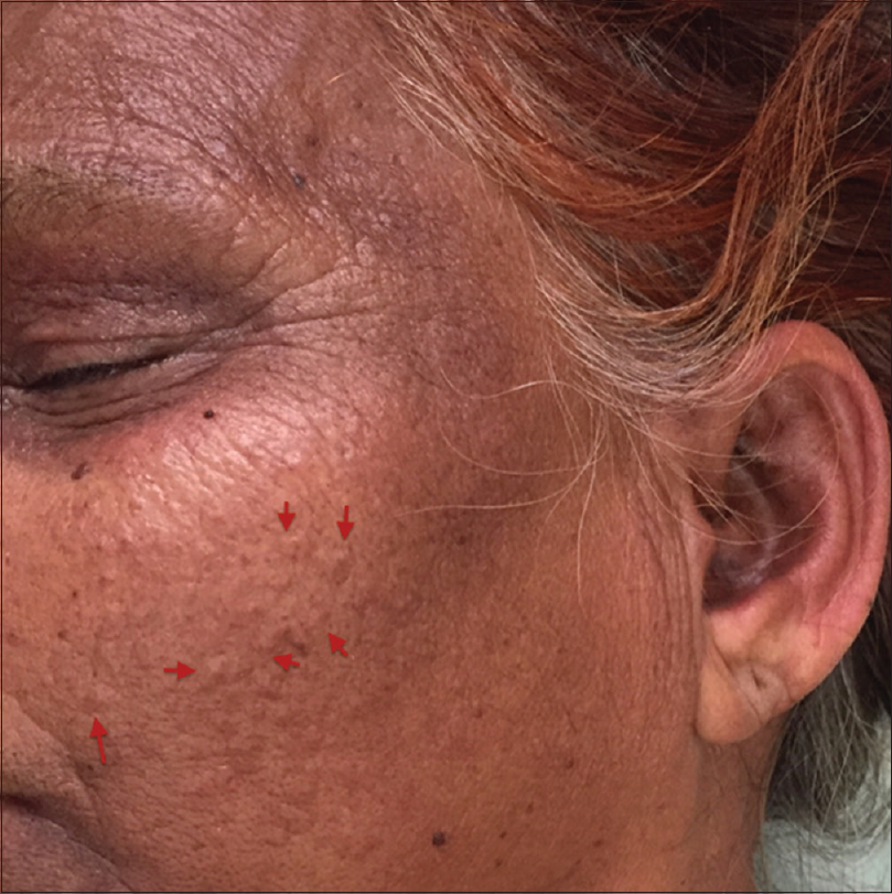
A 55-year-old woman, recently diagnosed with dermatomyositis, presented with multiple, tiny, firm papular lesions on cheeks for many years. These lesions were asymptomatic with no history of ulceration or discharge. Past history was notable for the presence of severe facial acne vulgaris during adolescence and early adulthood. On examination, there were multiple small round to irregular-shaped skin-colored, hard, monomorphic papules on both cheeks [Figure - 1]. There were no similar lesions elsewhere. Patient had characteristic cutaneous manifestations of dermatomyositis - V sign, shawl sign, heliotrope rash, Gottron's sign and holster sign) with proximal muscle weakness.
 |
| Figure 1: Multiple, round to irregularly shaped skin colored, monomorphic papules on cheek |
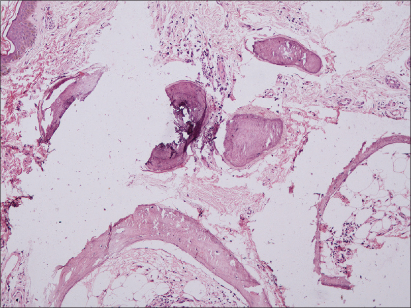
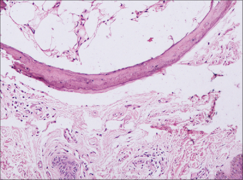
Histopathology from the papule showed fibrocollagenous tissue with round to irregular, large homogeneous amorphous basophilic staining deposits in the deep dermis and subcutis. The dermis also showed spicules of crescentic eosinophilic material housing cells held within small lacunae, associated with mature adipose tissue.[Figure - 2],[Figure - 3],[Figure - 4].
 |
| Figure 2: Round to irregular, large homogeneous amorphous basophilic staining deposits in the deep dermis and subcutis and spicules of crescentic eosinophilic material (H and E, 10x) |
 |
| Figure 3: Round to irregular, large homogeneous amorphous basophilic staining deposits in the deep dermis and subcutis and spicules of crescentic eosinophilic material (H and E, ×100) |
 |
| Figure 4: Crescentic eosinophilic material housing cells held within small lacunae, associated with mature adipose tissue (H and E, ×400) |
Question
What is your diagnosis?
| 1. |
Essing M. Osteoma cutis of the forehead. HNO 1985;33:548-50.
[Google Scholar]
|
| 2. |
Gfesser M, Worret WI, Hein R, Ring J. Multiple primary osteoma cutis. Arch Dermatol 1998;134:641-3.
[Google Scholar]
|
| 3. |
Mast AM, Hansen R. Multiple papules on the elbows. Congenital osteoma cutis. Arch Dermatol 1997;133:777-80.
[Google Scholar]
|
| 4. |
Altman JF, Nehal KS, Busam KJ, Halpern AC. Treatment of primary miliary osteoma cutis with incision, curettage, and primary closure. J Am Acad Dermatol 2001;44:96-9.
[Google Scholar]
|
| 5. |
MyllyläRM, Haapasaari KM, Palatsi R, Germain-Lee EL, Hägg PM, Ignatius J, et al. Multiple miliary osteoma cutis is a distinct disease entity: Four case reports and review of the literature. Br J Dermatol 2011;164:544-52.
[Google Scholar]
|
Fulltext Views
2,825
PDF downloads
2,140


