
Translate this page into:
Multiple proliferating trichilemmal cysts with cutaneous horn on scalp
Corresponding Author: Dr. Yichen Tang, Shanghai Skin Disease Hospital, 1278 Baode Road, Shanghai, China. tangyichen@medmail.com.cn
-
Received: ,
Accepted: ,
How to cite this article: Wu L, Jiang L, Tang Y. Multiple proliferating trichilemmal cysts with cutaneous horn on scalp. Indian J Dermatol Venereol Leprol 2023;89:765
A 45-year-old man was referred to our outpatient clinic with multiple soft skin tumours on the scalp, which had slowly grown for last 20 years. One year ago, a firm, nail-like keratin mass was found at the edge of the middle tumour [Figure 1]. The tumours were removed by radical excision surgery and histopathologically diagnosed as proliferating trichilemmal cyst [Figure 2].
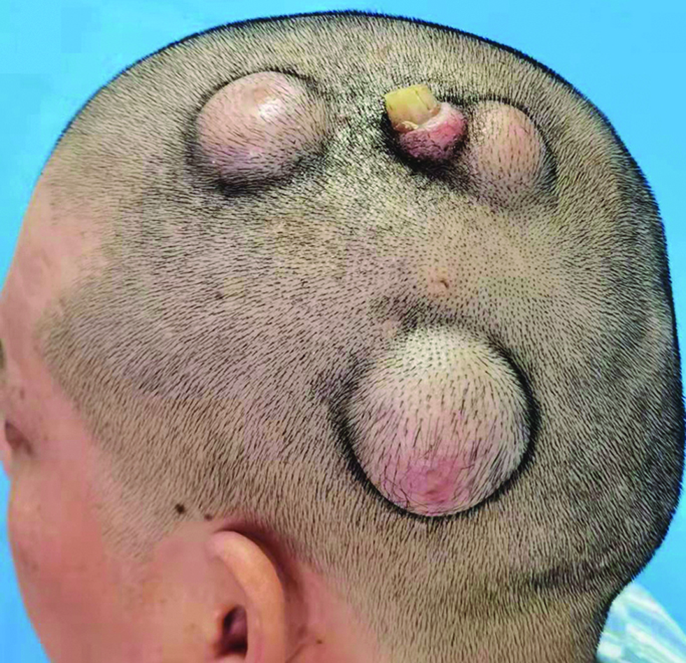
- Multiple skin tumours on the scalp with the middle tumour showing firm, nail-like keratin mass
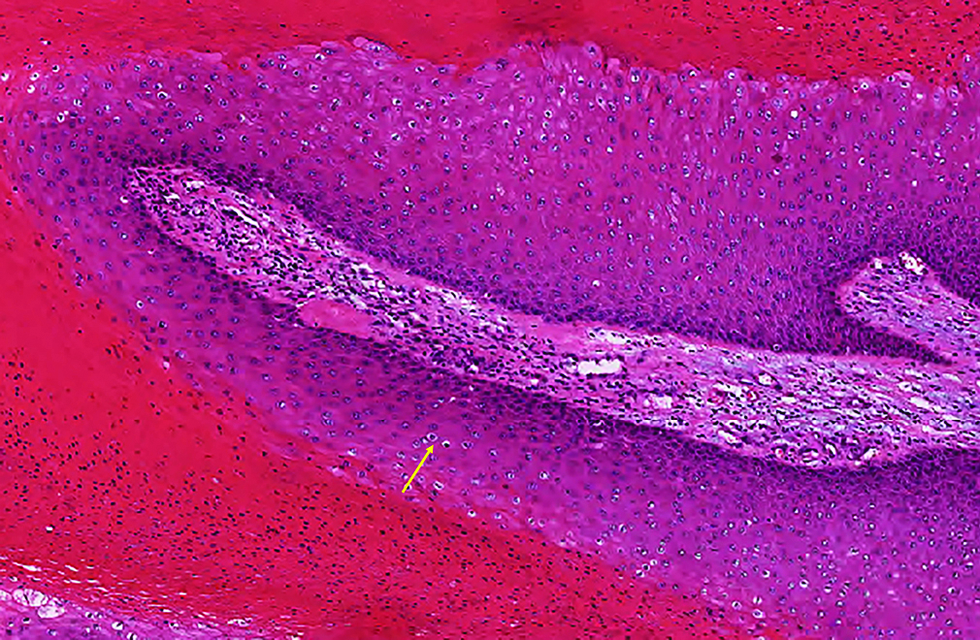
- Hematoxylin and eosin stained section showing a cyst in the dermis with the cyst wall composed of squamous epithelium without granular layer. The cells near the cyst cavity are large with the cytoplasm lightly stained (yellow arrow). Red-stained and densely arranged keratin is seen in the cyst. (H&E, ×200)
Association with human papillomavirus infection has been suspected in the pathogenesis of proliferating trichilemmal cysts. The histopathology of the tumour showed suspicious hollow cells in the local hyperplastic cyst wall, supporting a viral etiology. After the radical resection, there has been no recurrence or metastasis in this patient thus far.
Declaration of patient consent
The authors certify that they have obtained all appropriate patient consent forms.
Financial support and sponsorship
Nil.
Conflicts of interest
There are no conflicts of interest.


