Translate this page into:
Multiple ungual fibromas as an only cutaneous manifestation of tuberous sclerosis complex
2 T.R Ministry of Health Kecioren Education and Research Hospital, Ankara, Turkey
3 Department of Orthopaedics and Traumatology, Dıskapı Yıldırım Beyaz?t Education and Research Hospital, Ankara, Turkey
Correspondence Address:
Ezgi Unlu
Department of Dermatology, Zekai Tahir Burak Women's Health Education and Research Hospital, Samanpazarı-Ankara
Turkey
| How to cite this article: Unlu E, Balta I, Unlu S. Multiple ungual fibromas as an only cutaneous manifestation of tuberous sclerosis complex . Indian J Dermatol Venereol Leprol 2014;80:464-465 |
Sir,
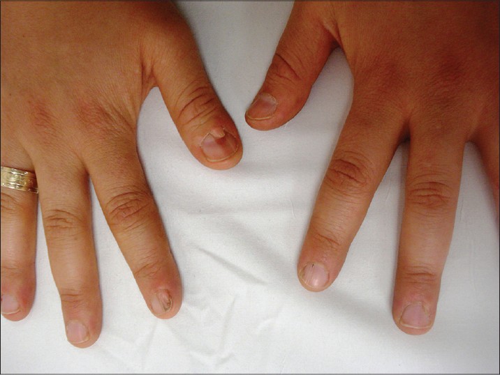
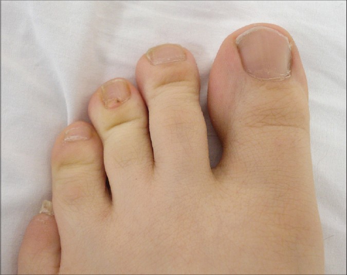
A 20-year-old woman presented to us with multiple firm, skin-colored lesions around two fingernails, one thumbnail and one toenail [Figure - 1] and [Figure - 2]. These asymptomatic lesions had appeared 8 years ago. She had initially visited the orthopaedics and traumatology department and they had referred her to our clinic for the diagnosis of these periungual lesions. As her skin lesions were quite classical, the diagnosis of ungual fibromas was made on clinical grounds without the requirement of histological confirmation. The patient was otherwise in good health and no other skin manifestation of tuberous sclerosis complex (TSC) was observed. Intraoral examination revealed multiple dental enamel pits. Gingival fibromas were not observed. Her family history was also negative for TSC. Examination of her eyes and cardiovascular system did not reveal any abnormality. Abdominal ultrasound imaging and X-rays of her hands and feet were within normal limits. Multiple subependymal nodules were detected by magnetic resonance imaging of the brain. The diagnosis of TSC was made on the basis of the above findings which fulfilled two major and one minor diagnostic criteria.
 |
| Figure 1: Ungual fibromas on fingernails and thumbnail |
 |
| Figure 2: An ungual fibroma on toenail |
She did not give consent for TSC1 and TSC2 gene analysis. Her family members were invited to our clinic for physical and radiological examinations but they declined to come. The patient was referred to the plastic surgery department for the treatment of her ungual fibromas.
Tuberous sclerosis is an autosomal dominant disorder characterized by multiple hamartomas of the skin, central nervous system, kidney, retina and heart. [1] It was first reported in 1880 by Bourneville who described a triad of epilepsy, mental deficiency and adenoma sebaceum. [2] Only 25% of the patients show the classical triad of the disease. The incidence of the disease is 1/10,000 and up to 50% of cases occur as a result of spontaneous mutations. [3]
The diagnosis of TSC can be made by genetic analysis or on the basis of clinical diagnostic criteria as given below. Three or more hypomelanotic macules with a size of at least 5 mm diameter, three or more facial angiofibromas or fibrous cephalic plaque, two or more ungual fibromas, shagreen patch, multiple retinal hamartomas, cortical dysplasias including tubers and cerebral white matter radial migration lines, subependymal nodules, subependymal giant cell astrocytoma, cardiac rhabdomyoma, lymphangioleiomyomatosis and two or more angiomyolipomas are the major criteria for the clinical diagnosis of TSC. Minor criteria include confetti skin lesions, three or more pits in dental enamel, two or more intraoral fibromas, retinal achromic patch, multiple renal cysts and non-renal hamartomas. Either two major criteria or one major criterion with two or more minor criteria are necessary for the definite diagnosis of TSC. In the 2012 International Tuberous Sclerosis Complex Consensus Conference, Northrup et al. suggested that a combination of lymphangioleiomyomatosis and angiomyolipomas without other features does not meet criteria for the definite diagnosis of TSC. Both of them together constitute only one major criterion. For possible diagnosis, either one major criterion or two or more minor criteria are needed. [4]
Multiple ungual fibromas are one of the major criteria for the diagnosis of TSC. [3] They appear as firm, smooth, skin-colored or reddish papules around fingernails and toenails. Ungual fibromas associated with TSC are known as Koenen′s tumors and are observed in 20% of patients. [4] Although, they usually appear after puberty, cases with onset in childhood have been reported. [3] They are usually asymptomatic but may sometimes cause nail deformity and pain.
Ungual fibromas are usually diagnosed while the patient is being examined for a different problem or if the lesions themselves are a cosmetic concern. They have typical clinical features and can usually be diagnosed on the basis of physical examination by dermatologists. If the lesion is not clinically obvious, histological examination is needed. Histological examination shows epidermal acanthosis and hyperkeratosis with a large number of dermal capillaries and fibroblasts.
The common loci of mutations causing TSC are in the TSC1 gene at 9q34 and TSC2 at 16p13.3 respectively. [3] Only 10% to 25% of patients do not show a pathogenic mutation of TSC1 and TSC2 genes by genetic analysis. While positive results have very high predictive value for family members, normal results do not exclude the diagnosis of TSC. [4] Unilateral facial angiofibromas, solitary cortical tubers and bilateral and multiple ungual fibromas have been described as segmental or oligosymptomatic forms of TSC. [5] Bilateral and multiple ungual fibromas have been reported as a result of cutaneous mosaicism. [5]
In conclusion, multiple ungual fibromas are important clues for the diagnosis of TSC and all cases must be carefully examined for other systemic manifestations of the disease. Visceral lesions may develop at a later point, so all patients should be asked to follow up regularly.
| 1. |
Berlin AL, Billick RC. Use of CO2 laser in the treatment of periungual fibromas associated with tuberous sclerosis. Dermatol Surg 2002;28:434-6.
[Google Scholar]
|
| 2. |
Hanno R, Beck R. Tuberous sclerosis. Neurol Clin 1987;5:351-60.
[Google Scholar]
|
| 3. |
Mazaira M, del Pozo Losada J, Fernández-Jorge B, Fernández-Torres R, Martínez W, Fonseca E. Shave and phenolization of periungual fibromas, Koenen's tumors, in a patient with tuberous sclerosis. Dermatol Surg 2008;34:111-3.
[Google Scholar]
|
| 4. |
Northrup H, Krueger DA. International Tuberous Sclerosis Complex Consensus Group. Tuberous sclerosis complex diagnostic criteria update: Recommendations of the 2012 International Tuberous Sclerosis Complex Consensus Conference. Pediatr Neurol 2013;49:243-54.
[Google Scholar]
|
| 5. |
Ruiz-Villaverde R, Blasco-Melguizo J, Hernández-Jurado I, Naranjo-Sintes R, Gutierrez Salmerón MT. Bilateral and multiple periungual fibromas as an oligosymptomatic form of tuberous sclerosis. Dermatology 2004;209:160-1.
[Google Scholar]
|
Fulltext Views
3,615
PDF downloads
2,058


