Translate this page into:
Mutilating basal cell carcinoma
Correspondence Address:
Sarabjit Kaur
House No. 401, Sector 14, Rohtak - 124 001, Haryana
India
| How to cite this article: Kaur S, Jindal N, Jain V K. Mutilating basal cell carcinoma . Indian J Dermatol Venereol Leprol 2014;80:431 |
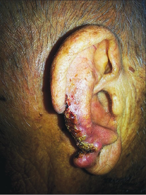
An elderly retired school teacher presented with a neglected ulcer over his right ear of 7 years duration. It began as a nodule and gradually eroded the ear lobe. At presentation, there was a large, deep mutilating ulcer measuring about 4 Χ 4 cm almost involving the helix of the right ear. The ulcer showed indurated edges, an erythematous base and a depressed floor with hemorrhagic, crusted and necrotic material [Figure - 1]. Telangiectasia was noted just above the ulcer. Histopathological examination showed a features suggestive of basal cell carcinoma. We referred him to the plastic surgeon for reconstructive surgery.
 |
| Figure 1: Mutilating basal cell carcinoma of the helix of the right ear |
Fulltext Views
1,803
PDF downloads
2,259


