Translate this page into:
Necrobiosis lipoidica mimicking cellulitis
Correspondence Address:
Neena Khanna
Department of Dermatology and Venereology, All India Institute of Medical Sciences, Ansari Nagar, New Delhi -110 029.
India
| How to cite this article: Joshi A, Rathi S K, Khanna N. Necrobiosis lipoidica mimicking cellulitis. Indian J Dermatol Venereol Leprol 1997;63:191-192 |
Abstract
A 57-year-old obese patient presented with a 5 month history of tender, indurated, erythematous plaques with superficial ulceration on the right shin. The lesions closely mimicked cellulitis but were unresponsive to antibiotics. Though the patient was not a known diabetic, on investigations she was found to be a diabetic. Histology confirmed the diagnosis of necrobiosis lipoidica. This acutely inflammed presentation of necrobiosis lipoidica is extremely rare. |
 |
Necrobiosis lipoidica (NL) is a degenerative disease of collagen with distinctive clinical presentation consisting of well-defined asymptomatic plaques of atrophic yellow skin which rarely ulcerate.[1][2] Necrobiosis lipoidica is a rare complication of diabetes mellitus (DM), occurring in about 0.3% of diabetic patients.[3][4] Between two-thirds to three-quarters of patients with NL have DM.[3][4] Although NL usually appears after DM, the two conditions can develop simultaneously and sometimes NL precedes DM.[5][6]We here report a case of NL with an unusual presentation.
Case Report
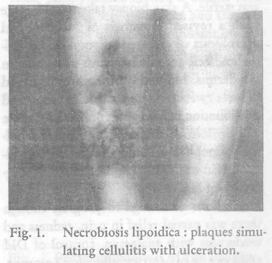
A 57-year-old obese, hypertensive female presented with a 5 month history of a deeply erythematous, moderately tender, and warm plaque on the right shin. There was no history suggestive of DM, nor was there a family history of DM. The patient had received courses of various systemic antibiotics and analgesics but the lesoin continued to rapidly progress and the plaque ulcerated. Other new lesions appeared within 4 months. Cutaneous examination revealed three irregular, ill-defined, intensely erythematous, tender, indurated 3x3 to 8xl0cm plaques. There was central depression and brown discoloration at the periphery of one lesion. A few, well-defined, punched-out small, round ulcers with necrotic base were present within the plaques. The lesions were not yellow or waxy. No telangiectasia were seen [Figure - 1]. An initial diagnosis of cellulitis was made and the patient was treated with courses of ampicillin, cloxacillin and erythromycin for 2 weeks each, but there was no change in the lesions.
Her fasting and post prandial blood sugar levels were found to be 196 mg/dl and 358 mg/dl respectively. The haemogram, renal and liver function tests were normal. Pus culture and sensitivity from the ulcerated lesion was sterile. A skin biopsy taken at this stage with a revised diagnosis of NL revealed necrobiosis of collagen surrounded by epithelioid cell granulomas with giant cells in the dermis. Marked proliferation of blood vessels with intimal thickening and hyalinisation of media was present. An acute and chronic inflammatory infiltrate including eosinophils extending upto the subcutaneous fat was seen. The features were suggestive of NL.
Her lesions continued to progress though there was partial relief in pain, redness and tenderness with analgesics, control of DM and a course of pentoxyfylline for 2 months.
Discussion
Necrobiosis lipoidica is a relatively rare condition associated with DM. Still rarer is the ulcerated and symptomatic form of NL. Both these features were present in our patient who did not have any past or family history of DM. Classical yellow-waxy appearance and telangiectases were missing and the patients lesions were rapidly progressive. The appearance simulated cellulitis and had resulted in patient being subjected to courses of antibiotics to which she was unresponsive. This prompted us to look for DM and do a skin biopsy which clinched the diagnosis of NL. The usual recalcitrant nature and relentless progression of the condition is well known. In our patient also the lesions continued to progress, though the lesions became less symptomatic after a course of analgesics and pentoxyfylline and on controlling her diabetes[7].
| 1. |
Hare PJ. Necrobiosis lipoidica, Br J Dermatol 1955;67:365-384.
[Google Scholar]
|
| 2. |
Olikarinen A, Mortenhomer M, Kallionen M et al. Necrobiosis lipoidica: Ultrastructural and biochemical demonstration of a collagen defect, J Invest Dermatol 1987;88:227-232.
[Google Scholar]
|
| 3. |
Hilderbrand AG, Montgomery H, Rynearson EH. Necrobiosis lipoidica diabeticorum, Arch Intern Med 1940;66:851-878.
[Google Scholar]
|
| 4. |
Gary HR, Graham JH, Johnson WC. Necrobiosis lipoidica: A histopathological and histochemical study, J Invest Dermatol 1965;44:369-380.
[Google Scholar]
|
| 5. |
Hill DM, Rhodes EL, Sheldon J et al, Necrobiosis lipoidica: Serum insulin and prednisone induced glycose tests in a non-diabetic group, Br J Dermatol 1966;78:332-336.
[Google Scholar]
|
| 6. |
Smith JG. Necrobiosis lipoidica: A disease of changing concepts, Arch Dermatol 1956;74:280-285.
[Google Scholar]
|
| 7. |
Engel MF, Hammack WJ. Necrobiosis lipoidica diabeticorum. A biochemical histochemical and electrophoretic study, Arch Dermatol 1958;78:73-81.
[Google Scholar]
|


