Translate this page into:
Normal hair follicle counts from scalp biopsy of Indian ethnicity: A cross-sectional study
Corresponding author: Dr. Sivaranjini Ramassamy, Department of Dermatology, Jawaharlal Institute of Postgraduate Medical Education and Research (JIPMER), Puducherry, India. siva11rsamy@yahoo.com
-
Received: ,
Accepted: ,
How to cite this article: Kololichalil A, Jinkala S, Ramassamy S. Normal hair follicle counts from scalp biopsy of Indian ethnicity: A cross-sectional study. Indian J Dermatol Venereol Leprol. 2025;91:111-2. doi: 10.25259/IJDVL_230_2024
Dear Editor,
Histological examination of scalp biopsy is the gold standard for the evaluation of alopecia. Biopsy specimens can be sectioned via the traditional/vertical approach or a horizontal/transverse approach. Horizontal section of scalp biopsy helps assess the density of hair follicles and morphometry of hair, especially in non-cicatricial alopecia.1 Normal follicle counts in the population are required for such interpretations. Hair density varies across ethnicities and there is heterogeneity among Asians.2 Studies have established reference data for hair follicle counts in Koreans, Thais, Iranians, and Taiwanese; quantitative data for Indians is lacking.2-5 Our objective was to determine the average hair count in the normal scalp of the south Indian Tamil population.
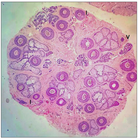
We obtained a 4 mm-punch biopsy from the occipital scalp of 40 subjects (17 men, 23 women). The participants were adults of south Indian ethnicity, defined as those residing in south India for the last three generations, aged 18 years and above, irrespective of gender, presenting for the evaluation of other hair disorders (in whom a normal scalp biopsy is indicated to interpret the pathology) and those undergoing follicular unit extraction as a part of vitiligo surgery or hair transplantation or scalp lesion excisions, who consented to give a 4 mm-punch sample from the occipital region. Those with a history of systemic diseases known to affect the hair cycle, those on topical or systemic medications affecting hair growth cycle within 6 months, known case of diffuse alopecia areata, clinically abnormal appearance of scalp and hair at the site of biopsy, positive hair pull test and abnormal dermoscopic findings at the site of scalp biopsy were excluded. The mean age of the participants was 29.50 years (range 18–54 years). We prepared the horizontal sections according to Whiting.1 Using standard definitions, follicular structures were identified and counted at the isthmic and sub-isthmic levels. The organisation of hair follicles into follicular units and appreciating the hair cycle morphology were possible at the above levels. The average number of follicular units and total hair in each 4mm biopsy were 8.07 and 21.38, respectively. The average terminal to vellus hair (T:V) and anagen to telogen (A:T) ratio were 9.3:1 and 95.05:4.95, respectively [Table 1 and Figure 1].
| Total (n=40) | Men (n=17) | Women (n=23) | p value | ≤19 years (n=7) | 20–39 years (n=25) | ≥40 years (n=8) | p value | |
|---|---|---|---|---|---|---|---|---|
| Age, mean±SD | 29.50±9.81 | 26.47±7.74 | 31.74±10.71 | 0.094 | - | - | - | - |
| Follicular units, mean±SD | 8.07±10.73 | 7.94±10.39 | 8.17±1.96 | 0.68 | 8.07±10.83 | 8.3±1.76 | 7.37±10.52 | 0.432 |
| Total hair, mean±SD | 21.38±5.99 | 19.14±3.53 | 22.78±6.98 | 0.057 | 24.28±6.06 | 20.74±5.36 | 20.12±7.64 | 0.332 |
| Follicular density/mm2, mean±SD | 1.69±00.47 | 1.52±00.28 | 1.81±0.55 | 0.057 | 1.93±00.48 | 1.64±00.42 | 1.61±00.60 | 0.332 |
| Terminal hair, mean±SD | 19.17±5.24 | 17.38±3.81 | 20.50±5.81 | 0.062 | 21.86±4.83 | 18.59±4.63 | 18.69±7.13 | 0.337 |
| Vellus hair, mean±SD | 2.06±10.72 | 1.73±10.19 | 2.3±2.03 | 0.056 | 2.43±10.74 | 2.16±10.82 | 1.44±10.42 | 0.497 |
| Terminal/vellus ratio | 9.3:1 | 10.04:1 | 8.91:1 | - | 9:1 | 8.6:1 | 12.98:1 | - |
| Anagen/Telogen ratio | 95.05:4.95 | 94.23:5.76 | 95.65:4.34 | 0.369 | 96.25:3.75 | 94.27:5.73 | 96.43:3.56 | 0.435 |
p value – statistical comparison between gender and age groups, SD- Standard deviation

- Horizontal section of occipital scalp biopsy at the isthmus level showing 9 follicular units, 23 total hair, 21 terminal hair, 1 vellus (V) hair, 2 intermediate (I) hair (1 each added to terminal and vellus count), 11:1 terminal vellus ratio and 100% hair in anagen in a 22-year-old man of South Indian ethnicity (Haematoxylin and eosin, 40×).
In the present study, the average number of follicular units was comparable to Koreans (7.8).3 It was slightly lower than other Asian populations of Thailand (9.1, 10.7 in different studies) and Taiwan (9.4) and much lower than Caucasians (14).2,4,5 The number of total hair was comparable to Thais (20.5), Taiwanese (21.3), and African Americans (21.5) but much lower than Iranians (36.4), Caucasians (35.5, 40 in different studies) and Hispanics (23.2).2-5 Among the Asians studied, the Koreans have a very low number of total hair (16.1) which may be explained by the inclusion of a few subjects with androgenetic alopecia (AGA) in their study.3 Normal scalp has a T:V ratio of >7:1 and anagen of at least 85%.1 These parameters were normal in our population and were similar to others, unaffected by ethnicity or race.
Our study had almost equal representation of men and women; we found no significant gender differences in hair parameters, except that the number of total hair was higher in women. This differed from the Thais and Taiwanese, where the number of total hair was higher in men.2,5 There was a decreasing trend in total hair and terminal hair count (not statistically significant) with the same number of follicular units from adolescents to older adults in our study. Our study sample had more representation of those aged 20–39 years; hence, it is unreliable to draw conclusions on the age differences in hair parameters.
The hair parameters found in our study could be compared with other studies, as the average age of the population studied was about 30 years in all of them. This study involves sampling from the occipital scalp which is androgen-insensitive and is least affected by miniaturisation. Thus, they represent a normal reference for hair density in the general population. The sample of 40 subjects was considered adequate to establish a mean for the studied population with the precision of 1 and a standard deviation of 3.2 at a 95% confidence interval, using the average terminal hairs in the Korean ethnicity reported by Lee et al. (14.9).3
To conclude, the present study explored hair count parameters in the normal south Indian Tamil population for interpreting horizontal sections of scalp biopsy from this ethnicity. We need studies from other ethnic groups within the country to get a national reference. Also, a trichoscopy and phototrichogram-guided calculation of hair follicle counts and morphometry analysis can be compared with the references obtained from histology as the former are less-invasive tools.
Ethical approval
The research/study was approved by the Institutional Review Board at JIPMER, Puducherry, number JIP/IEC/2021/002, dated 05-05-2021.
Declaration of patient consent
The authors certify that they have obtained all appropriate patient consent.
Financial support and sponsorship
Nil.
Conflicts of interest
There are no conflicts of interest.
Use of artificial intelligence (AI)-assisted technology for manuscript preparation
The authors confirm that there was no use of artificial intelligence (AI)-assisted technology for assisting in the writing or editing of the manuscript and no images were manipulated using AI.
References
- Diagnostic and predictive value of horizontal sections of scalp biopsy specimens in male pattern androgenetic alopecia. J Am Acad Dermatol. 1993;28:755-63.
- [CrossRef] [PubMed] [Google Scholar]
- The study of hair follicle counts from scalp histopathology in the Thai population. Int J Dermatol. 2020;59:978-81.
- [CrossRef] [PubMed] [Google Scholar]
- Hair counts from scalp biopsy specimens in Asians. J Am Acad Dermatol. 2002;46:218-21.
- [CrossRef] [PubMed] [Google Scholar]
- Hair counts in scalp biopsy of males and females with androgenetic alopecia compared with normal subjects. J Cutan Pathol. 2009;36:734-9.
- [CrossRef] [PubMed] [Google Scholar]
- Hair counts from normal scalp biopsy in taiwan. Dermatol Surg. 2012;38:1516-20.
- [CrossRef] [PubMed] [Google Scholar]


