Translate this page into:
Novel features in dermoscopy of lipoid proteinosis
Corresponding author: Dr. Ishan Agrawal, Postgraduate Resident, Department of Dermatology, IMS and SUM Hospital, Bhubaneswar, Odisha, India. ishanagrawal1995@gmail.com
-
Received: ,
Accepted: ,
How to cite this article: Ray A, Agrawal I, Singh BS, Kar BR. Novel features in dermoscopy of lipoid proteinosis. Indian J Dermatol Venereol Leprol 2023;89:116-8.
Sir,
Lipoid proteinosis is a rare progressive autosomal recessive disorder with infiltration of hyaline material in the skin, oral cavity, larynx and internal organs. Classically, the disease presents in early childhood with the initial manifestation being hoarseness of voice, however, late onset of the disease has been reported.1
A 23-year-old male, born to a non-consanguineous marriage, presented with a 6-month history of generalised pruritus and varied cutaneous lesions with the initial appearance of varioliform scars on the forehead. He also developed thickened verrucous plaques over bilateral elbows and on the gluteal cleft, multiple pinpoint papules over bilateral temporoparietal scalp and string of beaded papules on upper and lower palpebral margins [Figure 1a – c]. Oral mucosa was uninvolved. There were no other systemic complaints and no such complaints in the family. Our clinical diagnosis of lipoid proteinosis was confirmed by findings of periodic acid-Schiff (PAS)-positive homogenous hyaline deposits in the dermis on histopathology [Figure 2a and b].
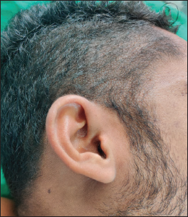
- Clinical presentation of Lipoid proteinosis (a) Pin-point papules on scalp
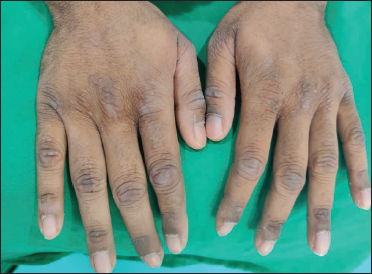
- Beaded papules with hyperpigmentation over dorsum of bilateral hands
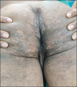
- Verrucous plaque, over intergluteal cleft
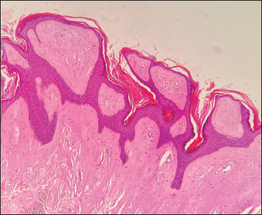
- Histopathology showing hyperkeratosis, papillomatosis and amorphous deposits in upper dermis (H and E, ×40)
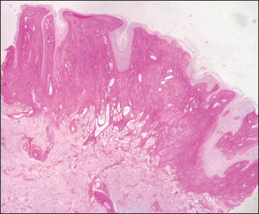
- PAS-positive deposition in upper dermis (PAS, ×40)
Dermoscopy of the five different areas, using a 3Gen Dermlite DL3 (CA, USA) dermoscope in polarised mode (×10) attached to an iPhoneX, showed varied features, as below:
Over the eyelids: The beaded appearance was prominent with clustered pale structureless globules in a linear configuration, along the upper and lower palpebral margin with misaligned eyelashes. Maralit et al. dermoscopically describe the lesions as whitish-yellow clod with brownish haloes.2
On the scalp: Multiple white-yellow structureless perifollicular globules without any loss of hair [Figure 3a].
Dorsum of hands: Multiple bluish-purple globules with scaling in the fissures and few haemorrhagic crusts [Figure 3b].
In the gluteal cleft: Multiple pearly, beaded globules clustered over the walls of the cleft with globules having centrally reddish-blue. Fine scales were visible over some areas [Figure 3c].
Over the elbows: Similar to Tabassum et al.’s dermoscopic findings, we observed yellow-red amorphous globules with slight scaling and multiple sulci and gyri, with a cerebriform appearance. The “pulpy” appearance described previously, was seen over the gluteal cleft and elbows in our case.3
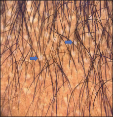
- Dermoscopy features: Multiple white-yellow, structureless, perifollicular dots (blue arrow) from the scalp (Dermlite DL3, ×10)
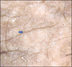
- Bluish-purple globules with scaling in fissures and hemorrhagic crust over the elbow (blue arrow), Dermlite DL3, ×10
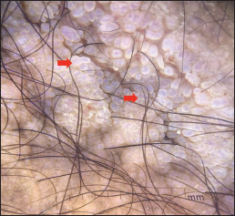
- Pearly beaded globules (red arrow) clustered over the intergluteal cleft, polarised mode (Dermlite DL3, ×10)
The structureless yellowish appearance of perifollicular and palpebral lesions is because of dermal deposition of eosinophilic hyaline amorphous material. Verrucous plaques in lipoid proteinosis are seen in areas where there is continued friction with perpendicularly oriented amorphous deposits to the epidermis.1 The scaling and cerebriform appearance of plaques is due to the underlying hyperkeratosis with papillomatosis and elongated rete ridges on histopathology.3
Deposition of hyaline material around epithelial adnexa including follicles causes patchy alopecia, however, in this case, dermoscopy showed intact follicles over amorphous globules on the scalp. This could be due to the relatively recent onset of the condition.1
Our findings highlight novel dermoscopic features of characteristic lesions in lipoid proteinosis, which have previously never been described.
Declaration of patient consent
A written consent was taken from the patient for the use of photographs.
Financial support and sponsorship
Nil.
Conflict of interest
There are no conflicts of interest.
References
- Lipoid proteinosis: A rare congenital genodermatosis. J NTR Univ Health Sci. 2017;6:166-8.
- [CrossRef] [Google Scholar]
- Lipoid proteinosis with esotropia: Report of a rare case and dermoscopic findings. Indian J Dermatol. 2020;65:53-6.
- [CrossRef] [PubMed] [Google Scholar]
- Clinincal, dermoscopy and histopathological findings in a case of lipoid proteinosis. J Phil Dermatol Soc. 2019;28:51-3.
- [Google Scholar]


